Hardware and
Instrumentation in
COMPUTED TOMOGRAPHY
Presented by: Dr. Anish Dhakal
Resident, MD Radiodiagnosis
KUSMS, Dhulikhel Hospital
27th
August, 2025
In a world before CT
Entire body areas were
inaccessible to radiography
- brain, mediastinum,
retroperitoneum
Diagnostic procedures
showing better detail in these
areas were potentially
harmful and or poorly
tolerated by the patient viz.
pneumoencephalography,
diagnostic
pneumomediastinum,
diagnostic laparotomy
Why CT?
Limitations of General Radiography:
• Superimposition of 3D structures over a 2D object
• Image is created directly on the image receptor
and is low in contrast because of Compton
scatter radiation
Historical Perspective
Godfrey Hounsfield (an engineer with Electric and Musical
Industries (EMI) Ltd. in England) is credited with the invention
of CT.
Early work on the mathematics used to reconstruct CT images:
Allan Cormack, a physicist at Tufts University
A first prototype of CT was installed at Atkinson Morley
Hospital under the aegis of Dr. James Ambrose in 1971 and its
clinical application started in 1972
The official announcement of invention of CT was made by G N
Hounsfield at the annual congress of British Institute of
Radiology in April 1972
The first body CT was installed by EMI at North Park Hospital in
1974
G N Hounsfield and Allan Cormack were awarded the 1979 Nobel
Prize for medicine for the invention of CT
First CT Scanner (EMI Mark I)
CT scanner in the early 1970s required 9 days to scan an object & produce a single-section
image.
Histological perspective
1973-74: The first II Generation CT system
total-body scanner has been realized in the U.S.,
the acquisition time for 1 tomogram was 18 sec.
1976-77: III and IV Generations CT with
acquisition time for 1 tomogram lower than 5 sec.
1983: First electron beam CT, very expensive.
1989: First helical CT, very low acquisition time
(less than 1 sec) and able to explore a large body
volume
2000: Multislice CT scanner, multiple arrays of
detectors; continuous development.
Introduction: Computed Tomography
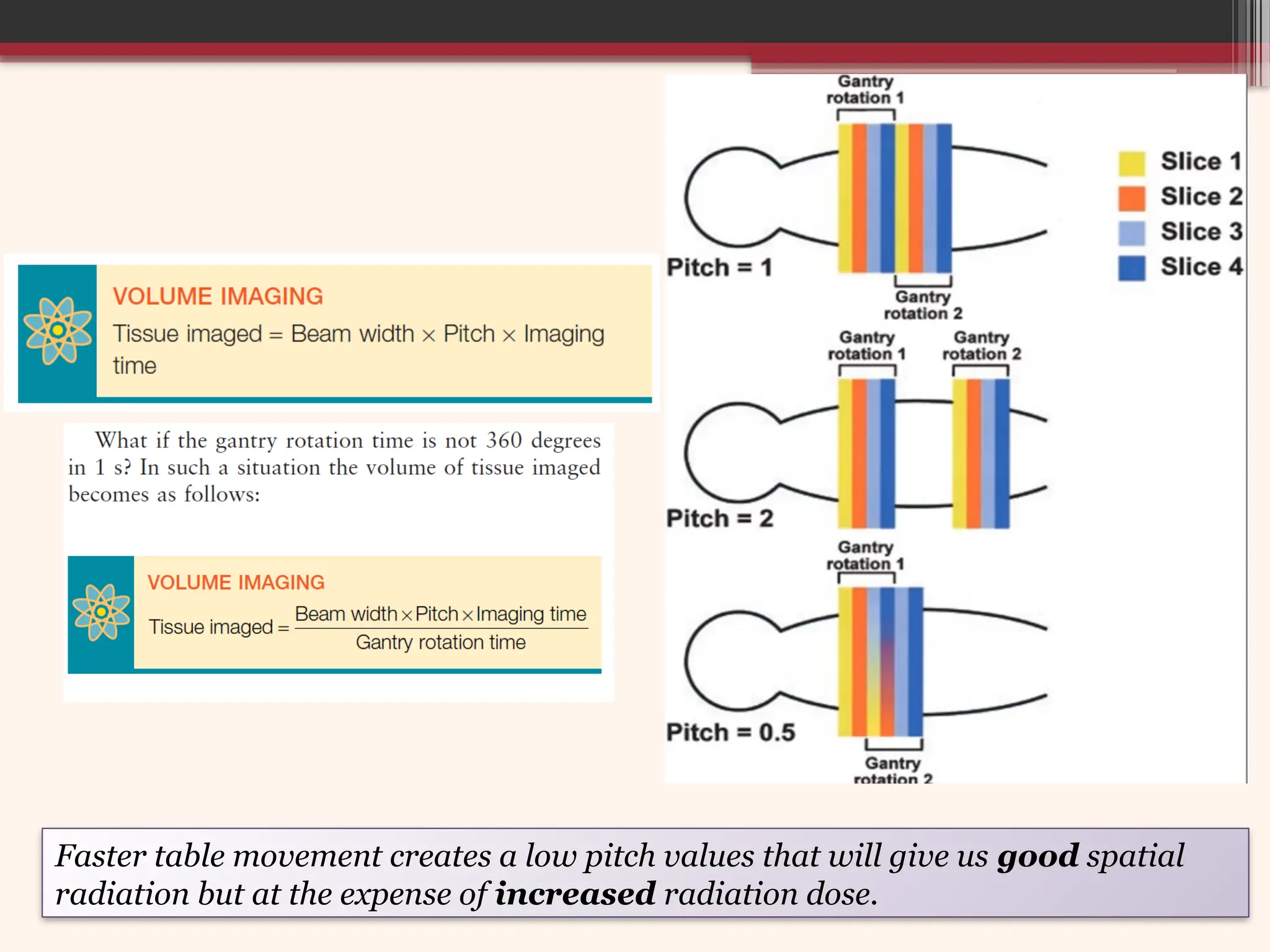
Greek words: Tomos (cut/slice/section) and Graphein
(write/record)
Literal definition: A form of tomography (imaging technique to
provide cross sectional images of the body) in which a computer
controls the motion of the X-ray source and detectors, processes the
data, and produces the image.
Computed Tomography is fundamentally a method of acquiring and
reconstructing an image of a thin cross section of an object
CT is a technique of creating tomographic images from digitized
data obtained by exposing the patient to x-rays from many different
angles.
Compared to radiographs, CT images are free of
superimposing tissues and are capable of much higher
contrast due to elimination of scatter.
BASIC PRINCIPLE OF CT
The internal structures of an object can be reconstructed
from multiple projection of the object
Basically, a narrow beam of X ray scans across a
patient in synchrony with an array of radiation detector
on the opposite side of the patient.
A sufficient no. of transmission measurements are taken
at different orientation of X ray source & detectors, the
distribution of attenuation coefficients within the layer
are determined
By assigning different gray levels to different
attenuation coefficients, an image can be reconstructed
with aid of computer that represent various structures
with different attenuation properties.
How CT differs from
conventional radiography?
CT scan differs from conventional projection in two significant
ways:
1. It forms a cross-sectional image, eliminating superimposition.
2. Sensitivity of CT to subtle differences in x-ray attenuation is at least
a factor of 10 higher than normally achieved by radiography, due to
the elimination of scatter.
The CT scan makes many measurements of attenuation through a
cross section.
Then these data are used to reconstruct a digital image of the cross
section, with each pixel in the image represent a measurement of
the mean attenuation of a volume element – voxel.
CT scanner Generations
Regardless of the CT scanner generation the latent
image is acquired and archived in a similar manner.
The exit radiation is detected & converted into a
digital signal by the analog-to-digital converter
(ADC).
Data from many different entrance angles are
processed in a computer to determine the
transmission & attenuation characteristics of the
tissues in the section under examination.
 Data are stored in a matrix of pixels. The digital
pixel data are processed in a digital-to-analog
converter (DAC) before being displayed.
What do CT generation mean?
Generation: Order in which CT scanner design
has been introduced, and each has a number
associated with it.
Classification based on arrangement of
components and mechanical motion required to
collect data.
Higher generation number doesn’t necessarily
indicate higher performance system.
First Generation
Very first CT scans were performed on a first generation geometry
on a CT benchtop. In the benchtop systems patients were not
imaged but rather an object to be imaged is placed on a stage
that can rotate (i.e. like a slow and well calibrated record player)
 Was a single ray system designed to examine only head.
 NaI scintillation detectors (one or two) with photomultiplier tubes
 X-ray beam: Finely collimated pencil thin slit field. X-ray beam turned on
while scanning and off during rotation.
 Tube rotation 180 degrees, Translate-rotate system.
 X-ray tube and two detectors connected in a C-arm fashion to move
synchronously from one side to other while scanning (translation). The x-ray
tube then rotates 1 degree into the next position and scans again.
 This process was performed 180 times for each scan.
 Nearly 5 minutes to complete one slice.
Use of two detectors split the finely collimated x-ray beam so that two contiguous
slices could be imaged during each procedure.
Early detector system couldn’t accommodate large
change in signal so patient head was recessed via a
rubber membrane into a water filled box/water
bath.
Acts to bolus the x-rays so that the intensity
outside the head is similar to the intensity inside
head .
Water bath allowed Hounsfield units (HU) to
maximize accuracy of attenuation coefficient
measurement (limitation of dynamic range, beam
hardening correction)
First generation CT
Advantage:
With regard to scatter rejection, pencil beam
geometry used in 1st
generation scanners were best.
Disadvantage:
Nearly 5 minutes was required to complete a
single image.
Contrast resolution of internal structures was
unprecedented, images had poor spatial resolution
Second Generation
Developed & installed by Ledley et al. at Georgetown
University in February 1974 (first waterless full body
scan)
Use a single projection fan-shaped beam Introduce
rest of the body part scanning, table movement
through, gantry angulation, & a laser indicator.
Translate-rotate system
Narrow fan beam (3-10 degrees) and multiple detector
elements (linear array of 30 detectors)
With 10 degree rotation increment, only 18 translation
required for 180 degrees image acquisition .
Shortest scan time: 18 sec/slice.
15 times faster than with 1st
generation CT.
Limitations in second
generation CT
Though speed improved, it was still limited by
mechanical complexity of translate – rotate geometry .
Even small deviations (because of vibration or other
misalignment) of scanner hardware position relative to
reconstruction voxels would cause data to be back
projected through wrong voxels creating severe artifacts.
Due to fan beam: Increased radiation intensity
towards the edge. Compensated with the use of bow-tie
filter (limits the range of intensity reaching detector and
hardens beam)
Third generation CT
The principal limitation of second-generation CT imaging systems
was examination time.
Because of the complex mechanical motion of translation and
rotation and the enormous mass involved in the gantry, most units
were designed for imaging times of 20 seconds or longer.
This limitation was overcome by third-generation CT imaging
systems.
Translation of source within each view was eliminated by
having a fan-beam shaped x-ray beam acquiring all the data (for a
slice) within each view by use of simple and pure rotational motion.
Accomplished by widening the x-ray beam encompassing the entire
patient width and using an array of detectors to intercept the beam.
With these imaging systems, the source and the detector array are
rotated about the patient.
As rotate-only units, third generation imaging systems can now
produce an image in less than 100 ms.
Third Generation
 Use a wider fan-shaped beam & a curved array of 250
to 750+ detectors.
 Rotate 360 degrees within the gantry.
 Rotate-rotate mechanism.
 Continues rotation of the detectors and x-ray tube in a circle
around the patient and the x-ray beam slices through the
body to produce image data.
 1 rotation, 1 second, 1 slice
 Solve the differential magnification problem which is caused
by the use of linear array of detectors, but problem of ring
artifacts.
The number of detectors and the width of the fan beam—
between 30 and 60 degrees—are both substantially larger
than for second-generation CT imaging systems.
In third generation CT imaging
systems the fan beam and the
detector array view the entire
patient at all times.
As the x-ray tube and detectors
rotate continuously, projection
profiles are collected and a view is
obtained for every fixed point of the
tube and detector and image is
reconstructed.
The curvilinear detector array
produces a constant source-to-
detector path length, which is an
advantage for good image
reconstruction (allows for better x-
ray beam collimation and reduces
the effect of scatter radiation)
• A finely collimated fan shaped x-
ray beam covering an angle of 30-
50 degree exposes the patient’s
body in many different angles
(about 1000) in a circular fashion
• The beam is accurately aligned to
an array of about 800-900 small
radiation detectors in the form of
an arc which measures the
transmitted intensity
• The measurements are digitized
and fed to a computer, which is
later reconstructed as
tomographic images of the body
part being exposed
Earlier 3rd generation CT:
Use of wraparound cable for gantry rotation.
Used Xenon detector arrange.
Xenon detector: Inherently stable and well matched because
factor affecting detector response were either uniform for the
entire array or constant over chamber.
Xenon detectors eventually replaced by solid state detectors.
Early third generation CT scanners installed on late 1975 could
scan in less than 5 sec ,
Modern variants of 3rd generation (MDCT):
Uses slip ring technology : K/a Continuously rotating fan beam
scanning.
Scan time: 0.2-0.5 sec.
Increases patient throughput
Limits the production of artifacts caused by respiratory motion
Disadvantage of 3rd
generation CT:
Requires extremely high detector stability
and matching of the detector response.
Any error or drift in the calibration of
detectors relative to other detectors result is
the ring artifact.
Sample size and spacing are fixed by detector
design
 Ring artifacts are never completely
eliminated , rather they are minimized by
high quality detectors design and frequent
calibration .
 Residual ring artifacts removed by image
processing algorithms.
 Despite these limitations , 3rd
generation CT
was highly successful (Slip Ring Technology)
and remains the basic geometry of most CT
scanners manufactured today.
Fourth Generation
Use a single projection fan shaped beam
with 600 to 2,000+ detectors mounted to an
array which forms a 360 degrees ring.
Rotate only movement, detectors remains
stationary
The tube first scans in a clockwise direction &
then counterclockwise; this motion continues
until the exam is complete.
1 minute for multiple slices.
Spatial resolution: >20 lp/cm.
Radiation detection is accomplished through a
fixed circular array of detectors, which contains as
many as 4000 individual detectors.
The x-ray beam is fan shaped with characteristics
similar to those of third-generation fan beams.
The fixed detector array of fourth-generation
CT imaging systems does not result in a constant
beam path from the source to all detectors, but it
does allow each detector to be calibrated and its
signal normalized for each image, as was possible
with second generation imaging systems.
Fourth-generation imaging systems were
developed because they were free of ring artifacts.
X-ray tube rotates outside
the detector ring
During rotation, the
detector ring tilts so that the
fan beam strikes an array of
detectors located at the far
side of the x-ray tube while
the detectors closest to the
x-ray tube move out of the
path of the x-ray beam.
Nutating: Tilting action of
the detector ring during
data collection.
Drawbacks:
Size and geometric dose inefficiency
Required large detector ring diameter.
Reduced spatial resolution limited detector aperture to
approx. 4 mm .
More scatter radiation, more patient dose. Scatter
absorbing septa used in 3rd
generation could not be used in
4th
generation because septa could necessarily be aimed at
center of the ring which was the source of scatter.
If such septa would used with stationary detectors they
would block primary x-rays from reaching the detectors at
oblique angles. Overcome by narrow collimation of x-ray
tube and software scatter correction.
Fourth Generation CT
Inside the geometry of 3rd
vs. 4th
generation CT
 3rd
generation: Fan beam geometry
has the x-ray tube as the apex of
the fan; 4th
generation has the
individual detector as the apex.
 3rd
generation-Detectors near the
edge of the detector array measure
the reference x-ray beam
 4th
generation-Reference beam is
measured by the same detector used
for transmission measurement
Flying focal spot (FFS) is used in Computed Tomography (CT) and other advanced X-ray imaging systems like
Digital Breast Tomosynthesis (DBT) to improve image quality by increasing sampling density and reducing
artifacts. By deflecting the X-ray focal spot, FFS technology enables the collection of more projection data with
a stationary detector, leading to higher spatial resolution, thinner slices, and the suppression of artifacts.
Fifth Generation
Is a dedicated cardiac unit designed around a
rotating electron beam. Also called the Electron
Beam CT scanner (EBCT).
Produces high-resolution images of moving organs
such as the (heart) without motion artifact.
The x-ray tube has been replaced with an electron gun
which uses deflection coils to direct a 30 degrees beam
of electrons in an arc around four adjacent tungsten
target rings (are stationary and span a 20-degree arc).
Ten times faster than conventional CT scanners, fast
enough to provide real-time dynamic sectional images
of the beating heart.
Why 5th
generation CT was
developed?
Cardiac imaging required ultra fast scan times (<50
ms) which was a hurdle with previous existed generation .
A novel CT scanner was developed specifically for cardiac
imaging which was capable of performing complete scans
in a little as 10-20 ms.
The idea behind the ultrafast scanner is a large bell
shaped x-ray tube.
Do not used conventional x-ray tube , instead a large arc
of tungsten encircles the patient and lies directly opposite
to the detector ring.
No moving parts in the gantry.
Electron beam is produced in cone like structures behind
the gantry and is electronically steered around the patient
so that it strikes the annular target.
Wherever it strikes – produces x-rays.
The concept is known as EBCT (Electron Beam CT)
Uses an electric gun that deflects & focuses the fast moving
electron beam along a 2100
arc of a large tungsten target
ring in the gantry.
When the focused electron beam scans this large target
area, X- rays are produced & collimated into a 2 cm wide
beam by a set of circular collimators.
X-ray beam traverses the patient & strikes the detector ring.
Two detector rings permits the simultaneous acquisition of
2 image sections.
Image of the whole heart can be acquired in ~0.2 s.
Although still available, EBCT was limited to cardiac screening mostly because of image
quality for general screening was lower than that of conventional CT (because of low mAs
values) and higher equipment costs. With progress being made cardiac scanning by multi
slice CT, the future of EBCT is uncertain.
Sixth Generation
 Generations one through four utilized electric cables to move the
components and to make X-ray exposure.
 Designed to use slip-ring technology to replace them.
 Allows continuous rotation of the x-ray tube & detectors
around the patient allowing for a continuous set of attenuation
data to be obtained in a helical/spiral manner (hence, helical/spiral
scanner).
 As the x-ray tube circles around the patient, the patient table is
continuously move through the gantry aperture.
 When the examination begins, the x-ray tube rotates continuously.
While the x-ray tube is rotating, the couch moves the patient through
the plane of the rotating x-ray beam. The x-ray tube is energized
continuously, data are collected continuously, and an image then can
be reconstructed at any desired z-axis position along the patient
3rd
and 4th
generation CT scanners eliminated the translate-
rotate motion , the gantry had to be stopped after each slice
was acquired.
Before Helical CT era: Cables are spooled onto a drum,
released during rotation and respooled during reversal.
Scanning, braking and reversal required at least 8-10 sec of
which only 1-2 sec were spent for data acquisition.
Results in poor temporal resolution and long procedure
time.
Three technological developments:
-Slip ring technology
-High power x-ray tubes
-Interpolation algorithms
After these improvements
Allowed true 3D image acquisition within a single
breath hold technique.
Patient is continuously translated while multiple
rotations of gantry (X ray tube and detector)
The path of x-ray tube and detector relative to the
patient is a helix.
An interpolation of the acquired measurement
data has to be performed in the z-direction to
estimate a complete CT data set at the desired
position.
Interpolation Algorithms
Reconstruction of an image at any z-axis position is possible
because of a mathematical process called interpolation.
If we estimate a value between known values, that is
interpolation; if one wishes to estimate a value beyond the
range of known values, that is extrapolation.
During helical CT, image data are received continuously and
when reconstructed, the plane of the image does not contain
enough data for reconstruction. Data in that plane must be
estimated by interpolation.
Data interpolation is performed by a special computer
program called an interpolation algorithm. The first
interpolation algorithms used 360-degree linear
interpolation.
The plane of the reconstructed
image was interpolated from data
acquired one revolution apart.
When these images are
formatted into sagittal and
coronal views, blurring can
occur. The solution to the
blurring problem is interpolation
of values separated by 180
degrees—half a revolution of the
x-ray tube.’
This results in improved z-axis
resolution and greatly improved
reformatted sagittal and coronal
views.
Allows production of additional
overlapping images with no
additional dose to the patient.
KNOWN
DATA
KNOWN
DATA
INTERPOLATE
D
DATA
Spiral scanning differs from
conventional CT scanning in that
the table is not stopped at the
center of each slice location
while the data are collected.
Advantage: CT examinations
time <1 minute, single breath-
hold examination, lower amount
of contrast media, decrease in
motion artifacts.
Acquisition time about 30
seconds, the acquisition time. and
the examination time are the
same.
 A conventional axial CT
examination requires several
minutes to complete, which is
much longer than a spiral CT
examination.
Advantages of sixth generation CT:
Fast scan times and large volume of data collected.
Minimizes motion artifacts.
Less misregistration between consecutive slices.
Reduced patient dose.
Improved spatial resolution.
Enhanced multiplaner or 3D renderings.
Improved temporal resolution.
Seventh generation CT (MS/MD
CT)
Multisection/Multislice/Multidetector CT (MSCT/MDCT).
Introduced in 1998.
Multiple rows of detectors-allows beam utilization.
Allows acquisition of multiple slice in single
rotation.
Faster scanning with a multiple row of detectors system
with multiple fan beams scanning simultaneously.
Large volume imaging possible with thin beams for
producing thin , high-detail slice images or 3-D images.
Minimum slice thickness- Minimum detector width not
by post patient collimator.
8, 16, 64, 128, 256, 320 and up to 640 slice CT
machine are available.
Able to expose multiple detectors
simultaneously due to detector technology
which permits an array of thousands of
parallel bands of detectors to operate at the
same time.
Coupled with helical scanning reduces the total
exam time for an entire chest or abdomen to 15 to
20 seconds.
Mathematically:
For e.g. A 128 slice CT refers to the number of detector rows (slices) the scanner can
acquire. Simultaneously per gantry rotation. If each slice is 0.625 mm, coverage per
rotation= 128x0.625 mm= 8 cm (wide Z-axis coverage)
Pitch: Table movement per rotation divided by beam width.
If beam width is 10mm , table moves 10 mm during one
tube rotation then pitch is 1 – x-ray beam associated with
consecutive helical loops are contiguous.
If beam width is 10 mm and table moves 15 mm per tube
rotation then pitch is 1.5 – gap exists between x-ray beam
edge of consecutive loop.
If beam width is 10 mm and table moves 7.5 mm then pitch
is 0.75 – beams and consecutive loops overlap by 2.5mm
(doubling exposure to the underlying tissues)
Increasing pitch to above 1 : 1 increases the volume of tissue
that can be imaged at a given time. This is one advantage of
multislice helical CT: the ability to image a larger volume of
tissue in a single breath-hold.
In practice, the pitch for multislice helical CT is usually 1.0.
Because multiple slices are obtained and z-axis location and
reconstruction width can be selected after imaging,
overlapping images are unnecessary.
An exception is CT angiography (CTA), which requires a
pitch of less than 1.0 : 1. Because of multislice capability,
more slices are acquired per unit time.
This results in a much larger volume of imaged tissue.
Higher pitch has faster coverage but reduced image quality
due to gaps in data.
Faster table movement creates a low pitch values that will give us good spatial
radiation but at the expense of increased radiation dose.
Components of a CT system
1. A gantry
2. A patient support
table/couch
3. A computer
system
4. An operator’s
console with
display
5. Accessories
1. The Gantry
Is a doughnut-shaped structure with X-ray
circuit, the x-ray tube, the radiation detectors,
the high-voltage generator, rotational
components including slip ring systems, gantry
angulation motors, patient positioning laser
lights and mechanical supports.
The gantry frame maintains the proper
alignment of the x-ray tube and detectors.
Gantry has a 50 to 80 cm aperture for the
patient to pass through during the scan.
Inside the gantry cover is a large ring that holds the
detectors and the track for the x-ray tube while it
rotates around the patient.
Can be angled up to 30 degrees toward or
away from the patient table to permit positioning
the patient for coronal images and to align the
slice plane to certain anatomy such as the base of
the skull or lumbar spine curvature.
Three intense white or low-power red laser lights:
used to accurately line the patient up for sagittal,
coronal, and transverse plane centering in the
aperture.
X-ray generator
Earlier: Low frequency (60 Hz), located outside the
gantry in the CT room & is connected to the rotating x-
ray tube by thick, flexible high-voltage cables.
The cables prevent the tube from rotating >360 degrees
without rewinding so axial CT examination collects data
one slice at a time.
Recent: High-frequency (3000 Hz) circuit, small
enough to be mounted with the x-ray tube on the
rotating frame inside the gantry.
Both the tube and the circuit rotate together
around the patient. The input voltage is connected to the
circuit through slip rings allowing the circuit and the x-
ray tube to continuously rotate.
X-Ray Filtration
Absorb soft, low energy X-
rays. Make uniformity of
X-ray spectrum.
Reduce scatter, reduce
patient dose
Improve image quality.
3 mm Al equivalent thickness
Flat: Copper or Aluminium.
Comb shaped/Bow-tie: Teflon
(a material with a low atomic
number & high density).
X-ray Tube
CT X-ray tube: Produces a continuous beam, high-heat
capacity tube, capable of operating up to 400 mA & 120 to
150 kVp for several seconds.
Heat storage capacity (3.5-7.5 MHU), designed to absorb
high heat levels generated from the high speed rotation of the
anode & the bombardment of electrons upon the anode
surface.
Exceed to a maximum heat value it will not operate until it
cools down to an acceptable level.
Anode: Larger diameter with graphite backing, which
allows the anode to absorb & dissipate large amounts of heat.
Focal spot size smaller (0.6 mm).
Bigger filament size, increased effective focal spot.
Anode target angle 7-10 degrees to diminish heel effect.
X-Ray Tube (Metal Ceramic type)
Glass envelope has been replaced by metal casing and
ceramic is used as insulation of high voltage cable e.g.-
super rotalix ceramic x-ray tube.
Metal envelope is grounded thus positive with respect to
electrons.
Anode rotates on an axle with bearing at each end providing
greater stability and reduced stress on shaft permits massive
anode approx-2 kg.
 Ceramic insulator (Al oxide) are used to insulate high
voltage parts of x-ray tube from metal envelope allowing more
compact tube design.
 Metal used is an alloy of chromium and iron.
 When metal enclosure is grounded to increase tube life.
 Higher tube loading, reduce off focus radiation, allow high tube
current.
X-Ray Tube (Metal Ceramic type)
Fig. Schematic diagram of a Super Rotalix ceramic x-ray tube: 1.Metal
casing, 2. Anode, 3/6. Ball bearings, 4/8. Ceramic insulators, 5. Cathode, 7. Stator
windings, 6. Anode shaft, 10. Beryllium window
X-Ray Tube
(Metal Ceramic type: Maximum rotalix)
Introduced Maximum rotalix ceramic (MRC)
tube by Philips-1989AD.
Features & advantage of MRC tube:
Improvement in rotating anode x-ray tube.
Noiselessly continuous rotating anode that
could be switched on in the morning and
switched off in the evening.
Liquid metal alloy as lubricant.
No waiting time during & between examination.
X-Ray Tube
(Metal Ceramic type: Maximum
rotalix)
MRC x-ray tube (Philips) with metal spiral groove
Possible to achieve dose saving filter technique in angiography.
Higher output & longer tube life.
Cont. rotation overcome the time to speed barrier.
200 mm graphite backed anode.
Anode heat storage capacity -8 MHU
Directly cooled Anode.
Tube voltage 90 to 140 kV.
Tube current 20 to 500 mA.
Anode angle – 7o .
Uses: Cardiovascular imaging, Multi-slice CT.
Metal Ceramic X-Ray Tube:
Aquilion Toshiba
High capacity multi-slice
CT tube
Heat storage capacity-7.5
MHU
Cooling rate- 1.7
MHU/min
Anode grounded
Focal spot 1.4 mm×1.6mm
Air cooled.
Aquilion x-ray tube
Straton X-Ray Tube
 Introduction of rotating envelope tube (RET)
 Idea was first documented in 1917.
 This design is very close to the realized Straton
tube (Siemens) having used magnetic &
deflecting coils to deflect (deflect focal spot 4,640
times) the electron beam for striking two areas
on the anode to get better z-axis resolution.
 Anode being direct contact with the cooling oil
enables an extremely high cooling rate (up
to 7 MHU /min).
 Made MDCT possible with improved workflow,
increased resolution.
 Focused and deflected beam of thermal electron
 Whole tube and anode assembly rotates.
 Bearings out side.
 Oil cooled, Zero heat storage capacity.
 Cooling rate- 4.7 MHU/min, Cooled down
within - 20 sec.
 Enables gantry speed of 0.37 seconds per
rotation.
 Tube current-500 mA.
In the early 1990s, the design of third- and fourth-
generation scanners evolved with the incorporation of a
new technology called slip ring.
A slip ring is a circular contact with sliding brushes which
supplies electrical power to the CT system and allows the
gantry to rotate continuously, uninterrupted by wires.
Electrical conductive rings and brushes transmit electrical
energy across a rotating interface.
The use of slip-ring technology eliminated the inertial
limitations at the end of each slice acquisition, and the
rotating gantry was free to rotate continuously
throughout the entire patient examination.
Slip ring Technology
Early CT- Used cables for power supply and data
transfer.
Leads to interscan delay.
No single breathhold examination possible.
There are usually 3 slip rings on a gantry:
 One provides high voltage power to the x-ray tube or low
voltage power to the high tension generator
 Second provides low voltage power to control systems on the
rotating gantry
 The third transfers digital data from the rotating detector array
Slip ring provides electrical
power to the rotating
components without fixed
connections.
Complete elimination of
interscan delays except for the
time required to move the
table to next slice position.
For eg : If scanning and
moving the table each take 1s ,
only 50% of the time is spent
acquiring the data .
Furthermore, rapid table
movement may introduce
tissue jiggle artifact.
Fig. Slip rings used to bring power to x-ray tube on rotating gantry of CT machine. (a) The shiny
metal strips carry electric signals that are swept off by special brushes. (b) The brushes are not
in the form of bristles but rather of metal blocks (in this case a silver alloy). The five pairs of
larger brushes provide the voltage required by the x-ray tube, and the three pairs of smaller ones
transfer signals from the gantry controller.
oDesigns:
DISC CYLINDER
BRUSH DESIGN: 2 Common Design
o Wire brush: Uses conductive wire as sliding contact
o Composite brush: Uses a block of some conductive
material ( e.g. silver alloy graphite ) as sliding contact
Based on the power supply:
1) Low voltage slip ring
• AC power and x-ray control signal transmitted to
slip rings by means of low voltage brushes that
glide in contact groove on the stationary slip ring.
• Slip rings then provide power to the high voltage
transformer which subsequently transmit to x-ray
tube and gantry.
AC SLIP RING
HV
GENERATO
R
X-
RAY
2) High voltage slip ring
AC delivers power to the high voltage generator which
subsequently supply high voltage to the slip ring.
High voltage from slip ring transmitted to x-ray tube.
In this design, high voltage generator does not rotate
with the x-ray tube
AC
HV
GENERATOR
SLIP
RING
X-RAY
Tube
Recent advance in the design of
slip rings: Contactless slip
ring.
Utilize IPT (Inductive power
transfer) to transfer power.
Electromagnetic field created
by coils placed in the stationery
transmitter and rotating
reciever.
Advantages of slip
ring technology
Contactless slip ring removes the inherent need for friction or contact to generate
an electrical current. Possible to transfer electrical energy across a rotating
interface without the use of electrical contacts. Reduces the maintenance costs.
•Removal of cable wrap around
process
•Elimination of the start–stop
process characteristic of conventional
CT scanners
•Provides capacity for continuous
data acquisition protocol like
dynamic studies and CTA.
•Faster scan time and minimal
interscan delay
Collimation
Collimation is required in CT scanning for exactly the same reasons as
in radiography.
Reduces patient dose by restricting the amount of tissue which is
irradiated.
Type of collimators:
 Pre-patient collimator (Tube or Source collimators):
 Is mounted on the tube housing and limits the area of the patient that
is exposed to the primary beam. This collimator determines the slice
thickness and patient dose.
 Post-patient collimator (Pre-detector or Anti-scatter collimators or
Anti-scatter septa or grid ):
 Is comprised of thin plates formed from a suitable X-ray absorbing
material like lead or tungsten.
Located directly below the patient & above the detectors restricts the
x-ray field viewed by the detector array.
Lead or Tungsten Plates are focused
at the x-ray focal spot and generally
located between columns of detectors
(z-direction) but not between rows of
detectors. This collimator is referred
to as a "1D" anti-scatter collimator.
In multi-slice scanners : shielding
between both columns & rows of
detectors; both directions are
focusing to the X-ray source. This
type is called a "2D" anti-scatter
collimator.
1-D Anti-scatter Collimator
2-D Anti-scatter Collimator
Radiation Detectors
 Detectors measure the intensity of radiation transmitted
through the patient.
Characteristics:
 Cost as minimum as possible.
 High absorption efficiency.
 High conversion efficiency.
 Responsiveness- Response time refers to length of time it
takes for a detector to review & discard signals.
 Dynamic range- Refers to the detector ability to receive wide
range of X ray intensities with proportional output. If exceed
dynamic range streak artifacts may occur.
Types of detectors:
 Gas ionization detectors
 Scintillation crystal detectors
Gas-filled Ionization Detectors
Use xenon gas to produce an electrical signal when x-
ray photons pass through the chamber.
Thin tungsten plates spaced 1.5 mm apart make up
the electrodes in the ionization chamber.
Space between the electrodes acts as radiation
detectors.
Each electrode produces an electric field across the
chamber. X-ray photons produce ionization in the
gas.
The electric field pulls the negative ions to the
positive electrode & the positive ions to the negative
electrode producing electric current.
Gas-filled Ionization Detectors
The current produced is proportional to the
ionization in the chamber and the energy of the
radiation passing through the chamber.
The detected energy makes up the digital signal that
is sent to the computer.
Detector efficiency approximately 50 - 60%.
Gas-filled Ionization Detectors
Diagram shows how an array of Xenon detector cells convert X-ray energy to the
digital data that is used by the image reconstruction computer.
Scintillation/Solid state Detectors
Comprised by sodium iodide crystals that give off a
flash of light when an x-ray photon is absorbed. Light is
produced in proportion to the intensity of the photon.
Use a Photomultiplier (PM) tube to convert x-ray photons
into an electrical signal which the computer uses to form
the visible image.
Light from the scintillation crystal is detected by a PM
tube. The light photon strikes the cathode of the PM tube
where it is converted into electrons.
Electrons are then amplified by a chain of dynodes as
they pass through the tube. The dynodes have
progressively higher voltage which causes an increase in
the number of electrons as they move toward the anode.
Once the electrons bombard the anode, they are
converted into an amplified electrical signal which is
then processed by the computer.
This system were used in early generations of scanners
but not to the modern scanners because of the
phosphorescent afterglow properties of them.
Sodium iodide was replaced by Bismuth germanate
& Cesium iodide.
The current crystal of choice is cadmium tungstate
which has more than 90% efficiency with minimal
afterglow.
Photodiode detects the light photons emitted, and
converts them into an electrical signal which is used by
the computer to form the digital image.
Scintiliation Detector
Diagram shows how an array of four Scintilating Crystal detector cells
convert X-ray energy to the digital data that is used by the image reconstruction computer.
2. Patient Support Table (Couch)
Either curved or flat.
Capable of moving up & down for ease in transferring
patients onto and off of the table, and in positioning the
patient correctly in the aperture.
Table indexing must be accurate and reproducible within
1 millimeter (mm).
Constructed of low atomic number carbon graphite
fiber to reduce attenuation of the x-ray beam and to
support patients weighing as much as 250-350 kg, if
excess may alter indexing .
Tabletop must be able to support the entire weight of the
patient when the table is moved into the gantry aperture.
In conventional CT scanning, the tube rotates
around the patient to collect data for one slice,
the table indexes into the gantry at a preset
distance, and the tube rotates again to collect
data for the next slice.
In helical scanning, table moves steadily through
the gantry while the tube continuously rotates
around the table and the patient.
The entire helical CT examination can be
completed in <1 minute.
3. Computer System
 Unique component of the CT system.
Sufficient speed & memory to solve several
thousands calculations simultaneously.
Is designed to control data acquisition,
processing, display, retrieve and storage.
Calculates the attenuation of the individual
voxels using algorithm.
Calculations of the CT numbers must be very
fast to produce images for immediate viewing.
4. Operator’s Console
Permits control of all scan parameters including
selecting proper technical factors, movement of the gantry
and patient table, and computer commands that allow
reconstruction and transfer of image data for storage in a
data file.
Operates a menu of directory operations.
Is preprogrammed with the kVp and mA values for
individual anatomic parts.
The technologist uses a keyboard and mouse to indicate the
desired operation for the anatomy to be scanned. Each scan
must have patient information, such as identification and
medical history entered prior to beginning the scan.
Hard copy images are printed on film using a multiformat
camera or printer.
Computed tomography
imaging systems can be
equipped with two or three
consoles. One console is used
by the CT technologist to
operate the imaging system.
Another console may be
available for a technologist to
postprocess images to annotate
patient data on the image (e.g.,
hospital identification, name,
patient number, age, gender)
and to provide identification
for each image (e.g., number,
technique, couch position).
Display console
Is often part of the main control and is a separate
CRT or flat panel display with controls.
May be a separate workstation that allows
radiologists to display and manipulate images,
and to electronically dictate the results, permit a
wide range of features to enhance the digital
image.
Multiplanar Reconstructions (MPRs), Reverse
Display, Magnification, Suppression, Selection of
Region of Interest (ROI), Annotation, Maximum
Intensity Projections (MIP), 3D Imaging etc.
5. ACCESSORIES
Head rests/supports, Knee support.
Velcro straps for immobilization.
Automatic contrast injector.
ECG gating machine for Cardiac gated CT.
Phantoms for BMD scanning.
Stereo-tactic localizer for brain lesion.
localization for stereo-tactic radio-surgery
planning.
Trolley with 2 compartments: sterilized and
unsterilized items.
Lead aprons, emergency drugs and
management system etc.
References
 Christensen's Physics of Diagnostic Radiology, 4th e
dition
 Radiologic Science For Technologists Physics Biolog
y And Protection by Stewart C. Bushong 12th edition
Thank you

Computed Tomography: Hardware and Instrumentation

  • 1.
    Hardware and Instrumentation in COMPUTEDTOMOGRAPHY Presented by: Dr. Anish Dhakal Resident, MD Radiodiagnosis KUSMS, Dhulikhel Hospital 27th August, 2025
  • 2.
    In a worldbefore CT Entire body areas were inaccessible to radiography - brain, mediastinum, retroperitoneum Diagnostic procedures showing better detail in these areas were potentially harmful and or poorly tolerated by the patient viz. pneumoencephalography, diagnostic pneumomediastinum, diagnostic laparotomy
  • 3.
    Why CT? Limitations ofGeneral Radiography: • Superimposition of 3D structures over a 2D object • Image is created directly on the image receptor and is low in contrast because of Compton scatter radiation
  • 4.
    Historical Perspective Godfrey Hounsfield(an engineer with Electric and Musical Industries (EMI) Ltd. in England) is credited with the invention of CT. Early work on the mathematics used to reconstruct CT images: Allan Cormack, a physicist at Tufts University A first prototype of CT was installed at Atkinson Morley Hospital under the aegis of Dr. James Ambrose in 1971 and its clinical application started in 1972 The official announcement of invention of CT was made by G N Hounsfield at the annual congress of British Institute of Radiology in April 1972 The first body CT was installed by EMI at North Park Hospital in 1974 G N Hounsfield and Allan Cormack were awarded the 1979 Nobel Prize for medicine for the invention of CT
  • 5.
    First CT Scanner(EMI Mark I) CT scanner in the early 1970s required 9 days to scan an object & produce a single-section image.
  • 6.
    Histological perspective 1973-74: Thefirst II Generation CT system total-body scanner has been realized in the U.S., the acquisition time for 1 tomogram was 18 sec. 1976-77: III and IV Generations CT with acquisition time for 1 tomogram lower than 5 sec. 1983: First electron beam CT, very expensive. 1989: First helical CT, very low acquisition time (less than 1 sec) and able to explore a large body volume 2000: Multislice CT scanner, multiple arrays of detectors; continuous development.
  • 7.
    Introduction: Computed Tomography Greekwords: Tomos (cut/slice/section) and Graphein (write/record) Literal definition: A form of tomography (imaging technique to provide cross sectional images of the body) in which a computer controls the motion of the X-ray source and detectors, processes the data, and produces the image. Computed Tomography is fundamentally a method of acquiring and reconstructing an image of a thin cross section of an object CT is a technique of creating tomographic images from digitized data obtained by exposing the patient to x-rays from many different angles. Compared to radiographs, CT images are free of superimposing tissues and are capable of much higher contrast due to elimination of scatter.
  • 8.
    BASIC PRINCIPLE OFCT The internal structures of an object can be reconstructed from multiple projection of the object Basically, a narrow beam of X ray scans across a patient in synchrony with an array of radiation detector on the opposite side of the patient. A sufficient no. of transmission measurements are taken at different orientation of X ray source & detectors, the distribution of attenuation coefficients within the layer are determined By assigning different gray levels to different attenuation coefficients, an image can be reconstructed with aid of computer that represent various structures with different attenuation properties.
  • 9.
    How CT differsfrom conventional radiography? CT scan differs from conventional projection in two significant ways: 1. It forms a cross-sectional image, eliminating superimposition. 2. Sensitivity of CT to subtle differences in x-ray attenuation is at least a factor of 10 higher than normally achieved by radiography, due to the elimination of scatter. The CT scan makes many measurements of attenuation through a cross section. Then these data are used to reconstruct a digital image of the cross section, with each pixel in the image represent a measurement of the mean attenuation of a volume element – voxel.
  • 14.
    CT scanner Generations Regardlessof the CT scanner generation the latent image is acquired and archived in a similar manner. The exit radiation is detected & converted into a digital signal by the analog-to-digital converter (ADC). Data from many different entrance angles are processed in a computer to determine the transmission & attenuation characteristics of the tissues in the section under examination.  Data are stored in a matrix of pixels. The digital pixel data are processed in a digital-to-analog converter (DAC) before being displayed.
  • 15.
    What do CTgeneration mean? Generation: Order in which CT scanner design has been introduced, and each has a number associated with it. Classification based on arrangement of components and mechanical motion required to collect data. Higher generation number doesn’t necessarily indicate higher performance system.
  • 16.
    First Generation Very firstCT scans were performed on a first generation geometry on a CT benchtop. In the benchtop systems patients were not imaged but rather an object to be imaged is placed on a stage that can rotate (i.e. like a slow and well calibrated record player)  Was a single ray system designed to examine only head.  NaI scintillation detectors (one or two) with photomultiplier tubes  X-ray beam: Finely collimated pencil thin slit field. X-ray beam turned on while scanning and off during rotation.  Tube rotation 180 degrees, Translate-rotate system.  X-ray tube and two detectors connected in a C-arm fashion to move synchronously from one side to other while scanning (translation). The x-ray tube then rotates 1 degree into the next position and scans again.  This process was performed 180 times for each scan.  Nearly 5 minutes to complete one slice. Use of two detectors split the finely collimated x-ray beam so that two contiguous slices could be imaged during each procedure.
  • 19.
    Early detector systemcouldn’t accommodate large change in signal so patient head was recessed via a rubber membrane into a water filled box/water bath. Acts to bolus the x-rays so that the intensity outside the head is similar to the intensity inside head . Water bath allowed Hounsfield units (HU) to maximize accuracy of attenuation coefficient measurement (limitation of dynamic range, beam hardening correction)
  • 20.
    First generation CT Advantage: Withregard to scatter rejection, pencil beam geometry used in 1st generation scanners were best. Disadvantage: Nearly 5 minutes was required to complete a single image. Contrast resolution of internal structures was unprecedented, images had poor spatial resolution
  • 21.
    Second Generation Developed &installed by Ledley et al. at Georgetown University in February 1974 (first waterless full body scan) Use a single projection fan-shaped beam Introduce rest of the body part scanning, table movement through, gantry angulation, & a laser indicator. Translate-rotate system Narrow fan beam (3-10 degrees) and multiple detector elements (linear array of 30 detectors) With 10 degree rotation increment, only 18 translation required for 180 degrees image acquisition . Shortest scan time: 18 sec/slice. 15 times faster than with 1st generation CT.
  • 23.
    Limitations in second generationCT Though speed improved, it was still limited by mechanical complexity of translate – rotate geometry . Even small deviations (because of vibration or other misalignment) of scanner hardware position relative to reconstruction voxels would cause data to be back projected through wrong voxels creating severe artifacts. Due to fan beam: Increased radiation intensity towards the edge. Compensated with the use of bow-tie filter (limits the range of intensity reaching detector and hardens beam)
  • 24.
    Third generation CT Theprincipal limitation of second-generation CT imaging systems was examination time. Because of the complex mechanical motion of translation and rotation and the enormous mass involved in the gantry, most units were designed for imaging times of 20 seconds or longer. This limitation was overcome by third-generation CT imaging systems. Translation of source within each view was eliminated by having a fan-beam shaped x-ray beam acquiring all the data (for a slice) within each view by use of simple and pure rotational motion. Accomplished by widening the x-ray beam encompassing the entire patient width and using an array of detectors to intercept the beam. With these imaging systems, the source and the detector array are rotated about the patient. As rotate-only units, third generation imaging systems can now produce an image in less than 100 ms.
  • 25.
    Third Generation  Usea wider fan-shaped beam & a curved array of 250 to 750+ detectors.  Rotate 360 degrees within the gantry.  Rotate-rotate mechanism.  Continues rotation of the detectors and x-ray tube in a circle around the patient and the x-ray beam slices through the body to produce image data.  1 rotation, 1 second, 1 slice  Solve the differential magnification problem which is caused by the use of linear array of detectors, but problem of ring artifacts. The number of detectors and the width of the fan beam— between 30 and 60 degrees—are both substantially larger than for second-generation CT imaging systems.
  • 26.
    In third generationCT imaging systems the fan beam and the detector array view the entire patient at all times. As the x-ray tube and detectors rotate continuously, projection profiles are collected and a view is obtained for every fixed point of the tube and detector and image is reconstructed. The curvilinear detector array produces a constant source-to- detector path length, which is an advantage for good image reconstruction (allows for better x- ray beam collimation and reduces the effect of scatter radiation)
  • 27.
    • A finelycollimated fan shaped x- ray beam covering an angle of 30- 50 degree exposes the patient’s body in many different angles (about 1000) in a circular fashion • The beam is accurately aligned to an array of about 800-900 small radiation detectors in the form of an arc which measures the transmitted intensity • The measurements are digitized and fed to a computer, which is later reconstructed as tomographic images of the body part being exposed
  • 28.
    Earlier 3rd generationCT: Use of wraparound cable for gantry rotation. Used Xenon detector arrange. Xenon detector: Inherently stable and well matched because factor affecting detector response were either uniform for the entire array or constant over chamber. Xenon detectors eventually replaced by solid state detectors. Early third generation CT scanners installed on late 1975 could scan in less than 5 sec , Modern variants of 3rd generation (MDCT): Uses slip ring technology : K/a Continuously rotating fan beam scanning. Scan time: 0.2-0.5 sec. Increases patient throughput Limits the production of artifacts caused by respiratory motion
  • 29.
    Disadvantage of 3rd generationCT: Requires extremely high detector stability and matching of the detector response. Any error or drift in the calibration of detectors relative to other detectors result is the ring artifact. Sample size and spacing are fixed by detector design  Ring artifacts are never completely eliminated , rather they are minimized by high quality detectors design and frequent calibration .  Residual ring artifacts removed by image processing algorithms.  Despite these limitations , 3rd generation CT was highly successful (Slip Ring Technology) and remains the basic geometry of most CT scanners manufactured today.
  • 30.
    Fourth Generation Use asingle projection fan shaped beam with 600 to 2,000+ detectors mounted to an array which forms a 360 degrees ring. Rotate only movement, detectors remains stationary The tube first scans in a clockwise direction & then counterclockwise; this motion continues until the exam is complete. 1 minute for multiple slices. Spatial resolution: >20 lp/cm.
  • 31.
    Radiation detection isaccomplished through a fixed circular array of detectors, which contains as many as 4000 individual detectors. The x-ray beam is fan shaped with characteristics similar to those of third-generation fan beams. The fixed detector array of fourth-generation CT imaging systems does not result in a constant beam path from the source to all detectors, but it does allow each detector to be calibrated and its signal normalized for each image, as was possible with second generation imaging systems. Fourth-generation imaging systems were developed because they were free of ring artifacts.
  • 33.
    X-ray tube rotatesoutside the detector ring During rotation, the detector ring tilts so that the fan beam strikes an array of detectors located at the far side of the x-ray tube while the detectors closest to the x-ray tube move out of the path of the x-ray beam. Nutating: Tilting action of the detector ring during data collection.
  • 34.
    Drawbacks: Size and geometricdose inefficiency Required large detector ring diameter. Reduced spatial resolution limited detector aperture to approx. 4 mm . More scatter radiation, more patient dose. Scatter absorbing septa used in 3rd generation could not be used in 4th generation because septa could necessarily be aimed at center of the ring which was the source of scatter. If such septa would used with stationary detectors they would block primary x-rays from reaching the detectors at oblique angles. Overcome by narrow collimation of x-ray tube and software scatter correction. Fourth Generation CT
  • 35.
    Inside the geometryof 3rd vs. 4th generation CT  3rd generation: Fan beam geometry has the x-ray tube as the apex of the fan; 4th generation has the individual detector as the apex.  3rd generation-Detectors near the edge of the detector array measure the reference x-ray beam  4th generation-Reference beam is measured by the same detector used for transmission measurement
  • 36.
    Flying focal spot(FFS) is used in Computed Tomography (CT) and other advanced X-ray imaging systems like Digital Breast Tomosynthesis (DBT) to improve image quality by increasing sampling density and reducing artifacts. By deflecting the X-ray focal spot, FFS technology enables the collection of more projection data with a stationary detector, leading to higher spatial resolution, thinner slices, and the suppression of artifacts.
  • 37.
    Fifth Generation Is adedicated cardiac unit designed around a rotating electron beam. Also called the Electron Beam CT scanner (EBCT). Produces high-resolution images of moving organs such as the (heart) without motion artifact. The x-ray tube has been replaced with an electron gun which uses deflection coils to direct a 30 degrees beam of electrons in an arc around four adjacent tungsten target rings (are stationary and span a 20-degree arc). Ten times faster than conventional CT scanners, fast enough to provide real-time dynamic sectional images of the beating heart.
  • 38.
    Why 5th generation CTwas developed? Cardiac imaging required ultra fast scan times (<50 ms) which was a hurdle with previous existed generation . A novel CT scanner was developed specifically for cardiac imaging which was capable of performing complete scans in a little as 10-20 ms. The idea behind the ultrafast scanner is a large bell shaped x-ray tube. Do not used conventional x-ray tube , instead a large arc of tungsten encircles the patient and lies directly opposite to the detector ring. No moving parts in the gantry.
  • 39.
    Electron beam isproduced in cone like structures behind the gantry and is electronically steered around the patient so that it strikes the annular target. Wherever it strikes – produces x-rays. The concept is known as EBCT (Electron Beam CT) Uses an electric gun that deflects & focuses the fast moving electron beam along a 2100 arc of a large tungsten target ring in the gantry. When the focused electron beam scans this large target area, X- rays are produced & collimated into a 2 cm wide beam by a set of circular collimators. X-ray beam traverses the patient & strikes the detector ring. Two detector rings permits the simultaneous acquisition of 2 image sections. Image of the whole heart can be acquired in ~0.2 s.
  • 40.
    Although still available,EBCT was limited to cardiac screening mostly because of image quality for general screening was lower than that of conventional CT (because of low mAs values) and higher equipment costs. With progress being made cardiac scanning by multi slice CT, the future of EBCT is uncertain.
  • 42.
    Sixth Generation  Generationsone through four utilized electric cables to move the components and to make X-ray exposure.  Designed to use slip-ring technology to replace them.  Allows continuous rotation of the x-ray tube & detectors around the patient allowing for a continuous set of attenuation data to be obtained in a helical/spiral manner (hence, helical/spiral scanner).  As the x-ray tube circles around the patient, the patient table is continuously move through the gantry aperture.  When the examination begins, the x-ray tube rotates continuously. While the x-ray tube is rotating, the couch moves the patient through the plane of the rotating x-ray beam. The x-ray tube is energized continuously, data are collected continuously, and an image then can be reconstructed at any desired z-axis position along the patient
  • 43.
    3rd and 4th generation CTscanners eliminated the translate- rotate motion , the gantry had to be stopped after each slice was acquired. Before Helical CT era: Cables are spooled onto a drum, released during rotation and respooled during reversal. Scanning, braking and reversal required at least 8-10 sec of which only 1-2 sec were spent for data acquisition. Results in poor temporal resolution and long procedure time. Three technological developments: -Slip ring technology -High power x-ray tubes -Interpolation algorithms
  • 44.
    After these improvements Allowedtrue 3D image acquisition within a single breath hold technique. Patient is continuously translated while multiple rotations of gantry (X ray tube and detector) The path of x-ray tube and detector relative to the patient is a helix. An interpolation of the acquired measurement data has to be performed in the z-direction to estimate a complete CT data set at the desired position.
  • 45.
    Interpolation Algorithms Reconstruction ofan image at any z-axis position is possible because of a mathematical process called interpolation. If we estimate a value between known values, that is interpolation; if one wishes to estimate a value beyond the range of known values, that is extrapolation. During helical CT, image data are received continuously and when reconstructed, the plane of the image does not contain enough data for reconstruction. Data in that plane must be estimated by interpolation. Data interpolation is performed by a special computer program called an interpolation algorithm. The first interpolation algorithms used 360-degree linear interpolation.
  • 46.
    The plane ofthe reconstructed image was interpolated from data acquired one revolution apart. When these images are formatted into sagittal and coronal views, blurring can occur. The solution to the blurring problem is interpolation of values separated by 180 degrees—half a revolution of the x-ray tube.’ This results in improved z-axis resolution and greatly improved reformatted sagittal and coronal views. Allows production of additional overlapping images with no additional dose to the patient.
  • 47.
  • 48.
    Spiral scanning differsfrom conventional CT scanning in that the table is not stopped at the center of each slice location while the data are collected. Advantage: CT examinations time <1 minute, single breath- hold examination, lower amount of contrast media, decrease in motion artifacts. Acquisition time about 30 seconds, the acquisition time. and the examination time are the same.  A conventional axial CT examination requires several minutes to complete, which is much longer than a spiral CT examination.
  • 49.
    Advantages of sixthgeneration CT: Fast scan times and large volume of data collected. Minimizes motion artifacts. Less misregistration between consecutive slices. Reduced patient dose. Improved spatial resolution. Enhanced multiplaner or 3D renderings. Improved temporal resolution.
  • 50.
    Seventh generation CT(MS/MD CT) Multisection/Multislice/Multidetector CT (MSCT/MDCT). Introduced in 1998. Multiple rows of detectors-allows beam utilization. Allows acquisition of multiple slice in single rotation. Faster scanning with a multiple row of detectors system with multiple fan beams scanning simultaneously. Large volume imaging possible with thin beams for producing thin , high-detail slice images or 3-D images. Minimum slice thickness- Minimum detector width not by post patient collimator.
  • 51.
    8, 16, 64,128, 256, 320 and up to 640 slice CT machine are available. Able to expose multiple detectors simultaneously due to detector technology which permits an array of thousands of parallel bands of detectors to operate at the same time. Coupled with helical scanning reduces the total exam time for an entire chest or abdomen to 15 to 20 seconds. Mathematically: For e.g. A 128 slice CT refers to the number of detector rows (slices) the scanner can acquire. Simultaneously per gantry rotation. If each slice is 0.625 mm, coverage per rotation= 128x0.625 mm= 8 cm (wide Z-axis coverage)
  • 52.
    Pitch: Table movementper rotation divided by beam width. If beam width is 10mm , table moves 10 mm during one tube rotation then pitch is 1 – x-ray beam associated with consecutive helical loops are contiguous. If beam width is 10 mm and table moves 15 mm per tube rotation then pitch is 1.5 – gap exists between x-ray beam edge of consecutive loop. If beam width is 10 mm and table moves 7.5 mm then pitch is 0.75 – beams and consecutive loops overlap by 2.5mm (doubling exposure to the underlying tissues)
  • 53.
    Increasing pitch toabove 1 : 1 increases the volume of tissue that can be imaged at a given time. This is one advantage of multislice helical CT: the ability to image a larger volume of tissue in a single breath-hold. In practice, the pitch for multislice helical CT is usually 1.0. Because multiple slices are obtained and z-axis location and reconstruction width can be selected after imaging, overlapping images are unnecessary. An exception is CT angiography (CTA), which requires a pitch of less than 1.0 : 1. Because of multislice capability, more slices are acquired per unit time. This results in a much larger volume of imaged tissue. Higher pitch has faster coverage but reduced image quality due to gaps in data.
  • 54.
    Faster table movementcreates a low pitch values that will give us good spatial radiation but at the expense of increased radiation dose.
  • 55.
    Components of aCT system 1. A gantry 2. A patient support table/couch 3. A computer system 4. An operator’s console with display 5. Accessories
  • 56.
    1. The Gantry Isa doughnut-shaped structure with X-ray circuit, the x-ray tube, the radiation detectors, the high-voltage generator, rotational components including slip ring systems, gantry angulation motors, patient positioning laser lights and mechanical supports. The gantry frame maintains the proper alignment of the x-ray tube and detectors. Gantry has a 50 to 80 cm aperture for the patient to pass through during the scan.
  • 57.
    Inside the gantrycover is a large ring that holds the detectors and the track for the x-ray tube while it rotates around the patient. Can be angled up to 30 degrees toward or away from the patient table to permit positioning the patient for coronal images and to align the slice plane to certain anatomy such as the base of the skull or lumbar spine curvature. Three intense white or low-power red laser lights: used to accurately line the patient up for sagittal, coronal, and transverse plane centering in the aperture.
  • 58.
    X-ray generator Earlier: Lowfrequency (60 Hz), located outside the gantry in the CT room & is connected to the rotating x- ray tube by thick, flexible high-voltage cables. The cables prevent the tube from rotating >360 degrees without rewinding so axial CT examination collects data one slice at a time. Recent: High-frequency (3000 Hz) circuit, small enough to be mounted with the x-ray tube on the rotating frame inside the gantry. Both the tube and the circuit rotate together around the patient. The input voltage is connected to the circuit through slip rings allowing the circuit and the x- ray tube to continuously rotate.
  • 59.
    X-Ray Filtration Absorb soft,low energy X- rays. Make uniformity of X-ray spectrum. Reduce scatter, reduce patient dose Improve image quality. 3 mm Al equivalent thickness Flat: Copper or Aluminium. Comb shaped/Bow-tie: Teflon (a material with a low atomic number & high density).
  • 60.
    X-ray Tube CT X-raytube: Produces a continuous beam, high-heat capacity tube, capable of operating up to 400 mA & 120 to 150 kVp for several seconds. Heat storage capacity (3.5-7.5 MHU), designed to absorb high heat levels generated from the high speed rotation of the anode & the bombardment of electrons upon the anode surface. Exceed to a maximum heat value it will not operate until it cools down to an acceptable level. Anode: Larger diameter with graphite backing, which allows the anode to absorb & dissipate large amounts of heat. Focal spot size smaller (0.6 mm). Bigger filament size, increased effective focal spot. Anode target angle 7-10 degrees to diminish heel effect.
  • 61.
    X-Ray Tube (MetalCeramic type) Glass envelope has been replaced by metal casing and ceramic is used as insulation of high voltage cable e.g.- super rotalix ceramic x-ray tube. Metal envelope is grounded thus positive with respect to electrons. Anode rotates on an axle with bearing at each end providing greater stability and reduced stress on shaft permits massive anode approx-2 kg.  Ceramic insulator (Al oxide) are used to insulate high voltage parts of x-ray tube from metal envelope allowing more compact tube design.  Metal used is an alloy of chromium and iron.  When metal enclosure is grounded to increase tube life.  Higher tube loading, reduce off focus radiation, allow high tube current.
  • 62.
    X-Ray Tube (MetalCeramic type) Fig. Schematic diagram of a Super Rotalix ceramic x-ray tube: 1.Metal casing, 2. Anode, 3/6. Ball bearings, 4/8. Ceramic insulators, 5. Cathode, 7. Stator windings, 6. Anode shaft, 10. Beryllium window
  • 63.
    X-Ray Tube (Metal Ceramictype: Maximum rotalix) Introduced Maximum rotalix ceramic (MRC) tube by Philips-1989AD. Features & advantage of MRC tube: Improvement in rotating anode x-ray tube. Noiselessly continuous rotating anode that could be switched on in the morning and switched off in the evening. Liquid metal alloy as lubricant. No waiting time during & between examination.
  • 64.
    X-Ray Tube (Metal Ceramictype: Maximum rotalix) MRC x-ray tube (Philips) with metal spiral groove Possible to achieve dose saving filter technique in angiography. Higher output & longer tube life. Cont. rotation overcome the time to speed barrier. 200 mm graphite backed anode. Anode heat storage capacity -8 MHU Directly cooled Anode. Tube voltage 90 to 140 kV. Tube current 20 to 500 mA. Anode angle – 7o . Uses: Cardiovascular imaging, Multi-slice CT.
  • 65.
    Metal Ceramic X-RayTube: Aquilion Toshiba High capacity multi-slice CT tube Heat storage capacity-7.5 MHU Cooling rate- 1.7 MHU/min Anode grounded Focal spot 1.4 mm×1.6mm Air cooled. Aquilion x-ray tube
  • 66.
    Straton X-Ray Tube Introduction of rotating envelope tube (RET)  Idea was first documented in 1917.  This design is very close to the realized Straton tube (Siemens) having used magnetic & deflecting coils to deflect (deflect focal spot 4,640 times) the electron beam for striking two areas on the anode to get better z-axis resolution.  Anode being direct contact with the cooling oil enables an extremely high cooling rate (up to 7 MHU /min).  Made MDCT possible with improved workflow, increased resolution.  Focused and deflected beam of thermal electron  Whole tube and anode assembly rotates.  Bearings out side.  Oil cooled, Zero heat storage capacity.  Cooling rate- 4.7 MHU/min, Cooled down within - 20 sec.  Enables gantry speed of 0.37 seconds per rotation.  Tube current-500 mA.
  • 67.
    In the early1990s, the design of third- and fourth- generation scanners evolved with the incorporation of a new technology called slip ring. A slip ring is a circular contact with sliding brushes which supplies electrical power to the CT system and allows the gantry to rotate continuously, uninterrupted by wires. Electrical conductive rings and brushes transmit electrical energy across a rotating interface. The use of slip-ring technology eliminated the inertial limitations at the end of each slice acquisition, and the rotating gantry was free to rotate continuously throughout the entire patient examination. Slip ring Technology
  • 68.
    Early CT- Usedcables for power supply and data transfer. Leads to interscan delay. No single breathhold examination possible.
  • 69.
    There are usually3 slip rings on a gantry:  One provides high voltage power to the x-ray tube or low voltage power to the high tension generator  Second provides low voltage power to control systems on the rotating gantry  The third transfers digital data from the rotating detector array
  • 70.
    Slip ring provideselectrical power to the rotating components without fixed connections. Complete elimination of interscan delays except for the time required to move the table to next slice position. For eg : If scanning and moving the table each take 1s , only 50% of the time is spent acquiring the data . Furthermore, rapid table movement may introduce tissue jiggle artifact.
  • 71.
    Fig. Slip ringsused to bring power to x-ray tube on rotating gantry of CT machine. (a) The shiny metal strips carry electric signals that are swept off by special brushes. (b) The brushes are not in the form of bristles but rather of metal blocks (in this case a silver alloy). The five pairs of larger brushes provide the voltage required by the x-ray tube, and the three pairs of smaller ones transfer signals from the gantry controller.
  • 72.
  • 73.
    BRUSH DESIGN: 2Common Design o Wire brush: Uses conductive wire as sliding contact o Composite brush: Uses a block of some conductive material ( e.g. silver alloy graphite ) as sliding contact
  • 74.
    Based on thepower supply: 1) Low voltage slip ring • AC power and x-ray control signal transmitted to slip rings by means of low voltage brushes that glide in contact groove on the stationary slip ring. • Slip rings then provide power to the high voltage transformer which subsequently transmit to x-ray tube and gantry. AC SLIP RING HV GENERATO R X- RAY
  • 75.
    2) High voltageslip ring AC delivers power to the high voltage generator which subsequently supply high voltage to the slip ring. High voltage from slip ring transmitted to x-ray tube. In this design, high voltage generator does not rotate with the x-ray tube AC HV GENERATOR SLIP RING X-RAY Tube
  • 76.
    Recent advance inthe design of slip rings: Contactless slip ring. Utilize IPT (Inductive power transfer) to transfer power. Electromagnetic field created by coils placed in the stationery transmitter and rotating reciever.
  • 77.
    Advantages of slip ringtechnology Contactless slip ring removes the inherent need for friction or contact to generate an electrical current. Possible to transfer electrical energy across a rotating interface without the use of electrical contacts. Reduces the maintenance costs. •Removal of cable wrap around process •Elimination of the start–stop process characteristic of conventional CT scanners •Provides capacity for continuous data acquisition protocol like dynamic studies and CTA. •Faster scan time and minimal interscan delay
  • 78.
    Collimation Collimation is requiredin CT scanning for exactly the same reasons as in radiography. Reduces patient dose by restricting the amount of tissue which is irradiated. Type of collimators:  Pre-patient collimator (Tube or Source collimators):  Is mounted on the tube housing and limits the area of the patient that is exposed to the primary beam. This collimator determines the slice thickness and patient dose.  Post-patient collimator (Pre-detector or Anti-scatter collimators or Anti-scatter septa or grid ):  Is comprised of thin plates formed from a suitable X-ray absorbing material like lead or tungsten. Located directly below the patient & above the detectors restricts the x-ray field viewed by the detector array.
  • 79.
    Lead or TungstenPlates are focused at the x-ray focal spot and generally located between columns of detectors (z-direction) but not between rows of detectors. This collimator is referred to as a "1D" anti-scatter collimator. In multi-slice scanners : shielding between both columns & rows of detectors; both directions are focusing to the X-ray source. This type is called a "2D" anti-scatter collimator. 1-D Anti-scatter Collimator 2-D Anti-scatter Collimator
  • 80.
    Radiation Detectors  Detectorsmeasure the intensity of radiation transmitted through the patient. Characteristics:  Cost as minimum as possible.  High absorption efficiency.  High conversion efficiency.  Responsiveness- Response time refers to length of time it takes for a detector to review & discard signals.  Dynamic range- Refers to the detector ability to receive wide range of X ray intensities with proportional output. If exceed dynamic range streak artifacts may occur. Types of detectors:  Gas ionization detectors  Scintillation crystal detectors
  • 81.
    Gas-filled Ionization Detectors Usexenon gas to produce an electrical signal when x- ray photons pass through the chamber. Thin tungsten plates spaced 1.5 mm apart make up the electrodes in the ionization chamber. Space between the electrodes acts as radiation detectors. Each electrode produces an electric field across the chamber. X-ray photons produce ionization in the gas. The electric field pulls the negative ions to the positive electrode & the positive ions to the negative electrode producing electric current.
  • 82.
    Gas-filled Ionization Detectors Thecurrent produced is proportional to the ionization in the chamber and the energy of the radiation passing through the chamber. The detected energy makes up the digital signal that is sent to the computer. Detector efficiency approximately 50 - 60%.
  • 83.
    Gas-filled Ionization Detectors Diagramshows how an array of Xenon detector cells convert X-ray energy to the digital data that is used by the image reconstruction computer.
  • 84.
    Scintillation/Solid state Detectors Comprisedby sodium iodide crystals that give off a flash of light when an x-ray photon is absorbed. Light is produced in proportion to the intensity of the photon. Use a Photomultiplier (PM) tube to convert x-ray photons into an electrical signal which the computer uses to form the visible image. Light from the scintillation crystal is detected by a PM tube. The light photon strikes the cathode of the PM tube where it is converted into electrons. Electrons are then amplified by a chain of dynodes as they pass through the tube. The dynodes have progressively higher voltage which causes an increase in the number of electrons as they move toward the anode.
  • 85.
    Once the electronsbombard the anode, they are converted into an amplified electrical signal which is then processed by the computer. This system were used in early generations of scanners but not to the modern scanners because of the phosphorescent afterglow properties of them. Sodium iodide was replaced by Bismuth germanate & Cesium iodide. The current crystal of choice is cadmium tungstate which has more than 90% efficiency with minimal afterglow. Photodiode detects the light photons emitted, and converts them into an electrical signal which is used by the computer to form the digital image.
  • 86.
    Scintiliation Detector Diagram showshow an array of four Scintilating Crystal detector cells convert X-ray energy to the digital data that is used by the image reconstruction computer.
  • 87.
    2. Patient SupportTable (Couch) Either curved or flat. Capable of moving up & down for ease in transferring patients onto and off of the table, and in positioning the patient correctly in the aperture. Table indexing must be accurate and reproducible within 1 millimeter (mm). Constructed of low atomic number carbon graphite fiber to reduce attenuation of the x-ray beam and to support patients weighing as much as 250-350 kg, if excess may alter indexing . Tabletop must be able to support the entire weight of the patient when the table is moved into the gantry aperture.
  • 88.
    In conventional CTscanning, the tube rotates around the patient to collect data for one slice, the table indexes into the gantry at a preset distance, and the tube rotates again to collect data for the next slice. In helical scanning, table moves steadily through the gantry while the tube continuously rotates around the table and the patient. The entire helical CT examination can be completed in <1 minute.
  • 89.
    3. Computer System Unique component of the CT system. Sufficient speed & memory to solve several thousands calculations simultaneously. Is designed to control data acquisition, processing, display, retrieve and storage. Calculates the attenuation of the individual voxels using algorithm. Calculations of the CT numbers must be very fast to produce images for immediate viewing.
  • 90.
    4. Operator’s Console Permitscontrol of all scan parameters including selecting proper technical factors, movement of the gantry and patient table, and computer commands that allow reconstruction and transfer of image data for storage in a data file. Operates a menu of directory operations. Is preprogrammed with the kVp and mA values for individual anatomic parts. The technologist uses a keyboard and mouse to indicate the desired operation for the anatomy to be scanned. Each scan must have patient information, such as identification and medical history entered prior to beginning the scan. Hard copy images are printed on film using a multiformat camera or printer.
  • 91.
    Computed tomography imaging systemscan be equipped with two or three consoles. One console is used by the CT technologist to operate the imaging system. Another console may be available for a technologist to postprocess images to annotate patient data on the image (e.g., hospital identification, name, patient number, age, gender) and to provide identification for each image (e.g., number, technique, couch position).
  • 92.
    Display console Is oftenpart of the main control and is a separate CRT or flat panel display with controls. May be a separate workstation that allows radiologists to display and manipulate images, and to electronically dictate the results, permit a wide range of features to enhance the digital image. Multiplanar Reconstructions (MPRs), Reverse Display, Magnification, Suppression, Selection of Region of Interest (ROI), Annotation, Maximum Intensity Projections (MIP), 3D Imaging etc.
  • 93.
    5. ACCESSORIES Head rests/supports,Knee support. Velcro straps for immobilization. Automatic contrast injector. ECG gating machine for Cardiac gated CT. Phantoms for BMD scanning. Stereo-tactic localizer for brain lesion. localization for stereo-tactic radio-surgery planning. Trolley with 2 compartments: sterilized and unsterilized items. Lead aprons, emergency drugs and management system etc.
  • 95.
    References  Christensen's Physicsof Diagnostic Radiology, 4th e dition  Radiologic Science For Technologists Physics Biolog y And Protection by Stewart C. Bushong 12th edition
  • 96.