. 2016 Dec;13(12):989-992.
doi: 10.1038/nmeth.4046. Epub 2016 Oct 31.

Simultaneous dual-color fluorescence lifetime imaging with novel red-shifted fluorescent proteins

Affiliations

Simultaneous dual-color fluorescence lifetime imaging with novel red-shifted fluorescent proteins

Tal Laviv et al. Nat Methods. 2016 Dec.

Abstract

We describe a red-shifted fluorescence resonance energy transfer (FRET) pair optimized for dual-color fluorescence lifetime imaging (FLIM). This pair utilizes a newly developed FRET donor, monomeric cyan-excitable red fluorescent protein (mCyRFP1), which has a large Stokes shift and a monoexponential fluorescence lifetime decay. When used together with EGFP-based biosensors, the new pair enables simultaneous imaging of the activities of two signaling molecules in single dendritic spines undergoing structural plasticity.


Conflict of interest statement

Competing financial interests

The authors declare no competing financial interests.

Figures

Figure 1
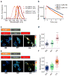
Figure 1. Characterization of CyRFP1 and dual-imaging with GCaMP6
(a) Normalized absorbance spectra of mCyRFP1, CyRFP1, and mEGFP. Dotted line indicates approximate 2p excitation (920 nm). (b) Normalized emission spectra of mCyRFP1, CyRFP1, and mEGFP. Dotted rectangles indicate transmission of green and red emission filters. (c) Fluorescence lifetime decay curves of coexpressed mEGFP and mCyRFP1 in HEK293 cells, fitted with a single exponential decay, alongside experimental instrument response function (IRF). (d) A dendritic segment of a CA1 pyramidal neuron in organotypic hippocampal slice transfected with CyRFP1 and GCaMP6s imaged with 2p microscopy. The spine indicated with the arrowhead was stimulated with 2p glutamate uncaging. Images before uncaging, 64 ms after the indicated number of uncaging pulses, and 30 s after the final uncaging are presented. (e) Quantification of changes in spine volume (Δ volume) and changes in the ratio of GCamp6 and CyRFP fluorescence (Δ ratio for) the spine showed in panel d during structural plasticity induced with glutamate uncaging. (f) In vivo 2p images of a dendritic segment of a layer 2/3 neuron in the motor cortex transfected with GCaMP6s and CyRFP1. The top panels show a spontaneous calcium elevation in spine 2. The bottom panels show a dendrite calcium transient. Merge, overlay of CyRFP1 and GCaMP6s images. (g) Timecourses of change in green-to-red fluorescence ratio in the two spines and the dendritic shaft in the image f. Inset shows time courses of CyRFP1 fluorescence. Scale bars, 2 μm.
Figure 2
Figure 2. Characterization of the mCyRFP1-mMaroon1 FRET pair for FLIM
(a) Normalized (norm.) spectra of absorptions and excitations (ex, excitation; em, emission) of mCyRFP1 and mMaroon1. (b) Fluorescence lifetime decay curves of dominant negative (DN) and constitutive active variants of a red-shifted RhoA sensor, RhoA–CyRM. (c) Schematics of a red-shifted Cdc42 sensor, Cdc42–CyRM, and representative fluorescence lifetime images of HEK293 cells expressing the sensor (WT) or the sensor with dominant negative (DN) or constitutively active (CA) mutations, acquired with 2pFLIM. (d) Binding fraction of Cdc42–CyRM mutants (80, 50, and 59 cells for DN, WT, and CA, respectively). (e) Schematics of RhoA–CyRM and representative fluorescence lifetime images of HEK293 cells expressing the sensor (WT) or its variants with DN or CA mutations. (f) Binding fraction of RyoA–CyRM mutants expressed in HEK293 cells (184, 190, and 181 cells for DN, WT, and CA, respectively). Scale bars, 20 μm. Error bars represent s.e.m.; statistical difference was measured using one-way ANOVA followed by Dunnett multiple comparison test.
Figure 3
Figure 3. Simultaneous FLIM measurements of RhoA and CaMKII activation in single dendritic spines undergoing structural plasticity
(a) Schematics of CaMKII-alpha sensor (Green-Camuiα) and RhoA–CyRM used for experiments. (b,c) Representative fluorescence lifetime images of RhoA (b) and CaMKII (c) activation in a dendritic segment of a CA1 pyramidal neuron in an organotypic hippocampal slice, acquired with 2pFLIM. Structural plasticity was induced in a single spine using 2p glutamate uncaging (marked with arrowhead). Scale bar, 2 μm. (d) Average changes in fluorescence lifetime of RhoA and CaMKII sensors during spine structural plasticity in the stimulated spines and their adjacent dendritic shafts (ten spines, eight neurons). (e) Quantifications of transient (top panel; 16–64 s for CaMKII and 16–76 s for RhoA) and sustained (bottom panel; 20–30 min) fluorescence lifetime changes in stimulated spines during simultaneous CaMKII–RhoA imaging under the following conditions: control, without extracellular Ca2+ (no Ca2+), replacing Green-Camuiα with mEGFP–dimVenus fusion (+mEGFP–dimVenus) and replacing acceptor of RhoA–CyRM with acceptor for Cdc42–CyRM (+RhoA false acceptor). (10/8, 10/8, 7/6, and 8/6 spines/cells, respectively; Error bars represent s.e.m.; statistical difference was measured using one-way ANOVA followed by Dunnett multiple comparison test).

References

    1. Jares-Erijman EA, Jovin TM. Nat Biotechnol. 2003:1387–1395. - PubMed
    1. Zhai S, Ark ED, Parra-Bueno P, Yasuda R. Science. 2013:1107–1111. - PMC - PubMed
    1. Murakoshi H, Wang H, Yasuda R. Nature. 2011;7:100–104. - PMC - PubMed
    1. Lee SJ, Escobedo-Lozoya Y, Szatmari EM, Yasuda R. Nature. 2009;58:299–304. - PMC - PubMed
    1. Nishiyama J, Yasuda R. Neuron. 2015;87:63–75. - PMC - PubMed

MeSH terms