. 2013 Apr;195(7):1360-70.
doi: 10.1128/JB.01989-12. Epub 2012 Nov 21.

The structure of the CS1 pilus of enterotoxigenic Escherichia coli reveals structural polymorphism

Affiliations

The structure of the CS1 pilus of enterotoxigenic Escherichia coli reveals structural polymorphism

Vitold E Galkin et al. J Bacteriol. 2013 Apr.

Abstract

Enterotoxigenic Escherichia coli (ETEC) is a bacterial pathogen that causes diarrhea in children and travelers in developing countries. ETEC adheres to host epithelial cells in the small intestine via a variety of different pili. The CS1 pilus is a prototype for a family of related pili, including the CFA/I pili, present on ETEC and other Gram-negative bacterial pathogens. These pili are assembled by an outer membrane usher protein that catalyzes subunit polymerization via donor strand complementation, in which the N terminus of each incoming pilin subunit fits into a hydrophobic groove in the terminal subunit, completing a β-sheet in the Ig fold. Here we determined a crystal structure of the CS1 major pilin subunit, CooA, to a 1.6-Å resolution. CooA is a globular protein with an Ig fold and is similar in structure to the CFA/I major pilin CfaB. We determined three distinct negative-stain electron microscopic reconstructions of the CS1 pilus and generated pseudoatomic-resolution pilus structures using the CooA crystal structure. CS1 pili adopt multiple structural states with differences in subunit orientations and packing. We propose that the structural perturbations are accommodated by flexibility in the N-terminal donor strand of CooA and by plasticity in interactions between exposed flexible loops on adjacent subunits. Our results suggest that CS1 and other pili of this class are extensible filaments that can be stretched in response to mechanical stress encountered during colonization.


Figures
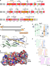
Fig 1
Fig 1
Crystal structure of ETEC CooAdsc. (A) Amino acid sequence alignment of CooA and CfaB. Secondary structure elements of CooA are indicated above its sequence. Red background, identical residues; yellow background, similar residues. (B) Schematic representation of wild-type CooA (top) and the CooAdsc construct (bottom). The sequence shown with orange background represents the Nte, which has been removed from the N terminus and attached to the C terminus. (C) Schematic of the secondary structure of CooAdsc. Green β-strands belong to the first β-sheet and blue and orange strands belong to the second β-sheet in the Ig fold. The orange G strand is the Nte, which has been fused to the C terminus. (D) Ribbon representation of the 1.6-Å-resolution CooAdsc crystal structure. (E) Space-filling representation of CooA with the Nte shown in stick representation. Carbon atoms are colored orange in the Nte, yellow in the DNKQ linker, and gray in the remainder of the protein. Oxygen atoms are red, and nitrogens are blue. Residues in the Nte are numbered as in native mature CooA, where the Nte of subunit n+1 (not shown) is donated to subunit n (shown). The Nte fits into the hydrophobic groove with hydrophobic side chains at positions P2 to P5, indicated in parentheses, filling corresponding pockets in the groove. (F) CS1 assembly scheme showing donor strand complementation between adjacent CooA subunits. Each subunit has a different color to illustrate donor strand complementation with an adjacent subunit in the filament.
Fig 2
Fig 2
Determination of the helical parameters of the CS1 pili. (A) Quick-freeze/deep-etch metal shadowed micrograph of CS1 pili and actin filaments. Scale bar, 1,000 Å. (Inset) Diffraction patterns obtained from F-actin (left) and CS1 pili (right). (B) Micrograph of the negatively stained CS1 filaments. Scale bar, 1,000 Å. (Inset) Diffraction pattern of negatively stained CS1 filaments reveals four major layer lines. Bessel orders and positions of the peaks are indicated.
Fig 3
Fig 3
IHRSR to determine CS1 symmetry. (A) IHRSR convergence of the overall set of CS1 segments from the three starting points yields two stable solutions. (B) IHRSR convergence of the three groups to their stable solutions starting from the same helical parameters.
Fig 4
Fig 4
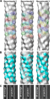
Characterization of three CS1 structural groups. (A) 3D reconstructions of the three structural groups (gray transparent surfaces) with the corresponding pseudoatomic-resolution models (colored ribbons). (B) Pseudoatomic models filtered to 20-Å resolution (cyan surfaces) superimposed on the 3D reconstructions (gray transparent surfaces). (C) Reference-free 2D averages of each structural group are compared with the projections of the corresponding 3D reconstructions.
Fig 5
Fig 5
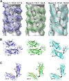
Comparison of the three CS1 structural states. (A) 3D reconstructions (Recon 1, 2, and 3) of the three structural states of the CS1 pili (transparent gray surfaces) shown with corresponding pseudoatomic models (ribbons). The reconstructions are contoured to show the head-to-tail connectivity between CooA subunits n and n+1 in the right-handed 1-start helix (dashed ovals). The Nte is shown in orange, and the N terminus of each CooAdsc subunit, Thr14, is shown as a black sphere. In the native CS1 filament, the Nte shown for subunit n is in fact the N-terminal strand of subunit n+1, linked via a peptide bond to Thr14 (indicated by arrows). (B) Orientation of and interactions between three subunits in the CS1 filament models, colored as in panel A. (C) Top view of subunits n and n+1 in the 1-start helix. The double-headed arrows indicate connectivity that would exist between Pro13 on the Nte bound to subunit n and Thr14 at the N terminus of subunit n+1.
Fig 6
Fig 6
Unraveling of CS1 pili. Quick-freeze/deep-etch metal shadowed micrograph of CS1 pili reveals an unraveling of helical filaments into fibrils at the ends and within CS1 filaments.

Comment in

References

    1. Qadri F, Svennerholm AM, Faruque AS, Sack RB. 2005. Enterotoxigenic Escherichia coli in developing countries: epidemiology, microbiology, clinical features, treatment, and prevention. Clin. Microbiol. Rev. 18:465–483 - PMC - PubMed
    1. Gaastra W, Svennerholm AM. 1996. Colonization factors of human enterotoxigenic Escherichia coli (ETEC). Trends Microbiol. 4:444–452 - PubMed
    1. Low D, Braaten B, Van de Woude M. 1996. Fimbriae, p 146–157 In Neidhardt RCIFC, Ingraham JL, Lin ECC, Low KB, Magasanik B, Reznikoff WS, Riley M, Schaechter M, Umbarger HE. (ed), Escherichia coli and Salmonella: cellular and molecular biology, vol 1 ASM Press, Washington, DC
    1. Sakellaris H, Scott JR. 1998. New tools in an old trade: CS1 pilus morphogenesis. Mol. Microbiol. 30:681–687 - PubMed
    1. Poole ST, McVeigh AL, Anantha RP, Lee LH, Akay YM, Pontzer EA, Scott DA, Bullitt E, Savarino SJ. 2007. Donor strand complementation governs intersubunit interaction of fimbriae of the alternate chaperone pathway. Mol. Microbiol. 63:1372–1384 - PubMed

Publication types

MeSH terms

LinkOut - more resources