Protein L: a novel reagent for the detection of chimeric antigen receptor (CAR) expression by flow cytometry
- PMID: 22330761
- PMCID: PMC3299624
- DOI: 10.1186/1479-5876-10-29
Protein L: a novel reagent for the detection of chimeric antigen receptor (CAR) expression by flow cytometry
Abstract
Background: There has been significant progress in the last two decades on the design of chimeric antigen receptors (CAR) for adoptive immunotherapy targeting tumor-associated antigens. Structurally CARs consist of a single chain antibody fragment directed against a tumor-associated antigen fused to an extracellular spacer and transmembrane domain followed by T cell cytoplasmic signaling moieties. Currently several clinical trials are underway using gene modified peripheral blood lymphocytes (PBL) with CARs directed against a variety of tumor associated antigens. Despite the improvements in the design of CARs and expansion of the number of target antigens, there is no universal flow cytometric method available to detect the expression of CARs on the surface of transduced lymphocytes.
Methods: Currently anti-fragment antigen binding (Fab) conjugates are most widely used to determine the expression of CARs on gene-modified lymphocytes by flow cytometry. The limitations of these reagents are that many of them are not commercially available, generally they are polyclonal antibodies and often the results are inconsistent. In an effort to develop a simple universal flow cytometric method to detect the expression of CARs, we employed protein L to determine the expression of CARs on transduced lymphocytes. Protein L is an immunoglobulin (Ig)-binding protein that binds to the variable light chains (kappa chain) of Ig without interfering with antigen binding site. Protein L binds to most classes of Ig and also binds to single-chain antibody fragments (scFv) and Fab fragments.
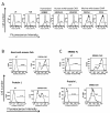
Results: We used CARs derived from both human and murine antibodies to validate this novel protein L based flow cytometric method and the results correlated well with other established methods. Activated human PBLs were transduced with retroviral vectors expressing two human antibody based CARs (anti-EGFRvIII, and anti-VEGFR2), two murine antibody derived CARs (anti-CSPG4, and anti-CD19), and two humanized mouse antibody based CARs (anti-ERBB2, and anti-PSCA). Transduced cells were stained first with biotin labeled protein L followed by phycoerythrin (PE)-conjugated streptavidin (SA) and analyzed by flow cytometry. For comparison, cells were stained in parallel with biotin conjugated goat-anti-mouse Fab or CAR specific fusion proteins. Using protein L, all CAR transduced lymphocytes exhibited specific staining pattern ranging from 40 to 80% of positive cells (compared to untransduced cells) and staining was comparable to the pattern observed with anti-Fab antibodies.
Conclusion: Our data demonstrate the feasibility of employing Protein L as a general reagent for the detection of CAR expression on transduced lymphocytes by flow cytometry.
Figures


References
-
- Zhao Y, Wang QJ, Yang S, Kochenderfer JN, Zheng Z, Zhong X, Sadelain M, Eshhar Z, Rosenberg SA, Morgan RA. A herceptin-based chimeric antigen receptor with modified signaling domains leads to enhanced survival of transduced T lymphocytes and antitumor activity. J Immunol. 2009;183:5563–5574. doi: 10.4049/jimmunol.0900447. - DOI - PMC - PubMed
MeSH terms
Substances
LinkOut - more resources
Full Text Sources
Other Literature Sources
Research Materials
Miscellaneous

