. 2009 May;191(9):3149-61.
doi: 10.1128/JB.01701-08. Epub 2009 Feb 27.

Docking and assembly of the type II secretion complex of Vibrio cholerae

Affiliations

Docking and assembly of the type II secretion complex of Vibrio cholerae

Suzanne R Lybarger et al. J Bacteriol. 2009 May.

Abstract

Secretion of cholera toxin and other virulence factors from Vibrio cholerae is mediated by the type II secretion (T2S) apparatus, a multiprotein complex composed of both inner and outer membrane proteins. To better understand the mechanism by which the T2S complex coordinates translocation of its substrates, we are examining the protein-protein interactions of its components, encoded by the extracellular protein secretion (eps) genes. In this study, we took a cell biological approach, observing the dynamics of fluorescently tagged EpsC and EpsM proteins in vivo. We report that the level and context of fluorescent protein fusion expression can have a bold effect on subcellular location and that chromosomal, intraoperon expression conditions are optimal for determining the intracellular locations of fusion proteins. Fluorescently tagged, chromosomally expressed EpsC and EpsM form discrete foci along the lengths of the cells, different from the polar localization for green fluorescent protein (GFP)-EpsM previously described, as the fusions are balanced with all their interacting partner proteins within the T2S complex. Additionally, we observed that fluorescent foci in both chromosomal GFP-EpsC- and GFP-EpsM-expressing strains disperse upon deletion of epsD, suggesting that EpsD is critical to the localization of EpsC and EpsM and perhaps their assembly into the T2S complex.


Figures

FIG. 1.
FIG. 1.
Distribution of GFP chimeras varies with expression level and context. Plasmid-borne GFP-EpsM in live cells of V. cholerae epsM mutant PU3 was polarly localized when overexpressed with 10 μM IPTG (A) but circumferentially distributed when not induced (B). Both patterns differed from that of chromosomally expressed GFP-EpsM, balanced with the other Eps proteins, which formed fluorescent foci along the cell membranes (C). (D) GFP-EpsC, expressed from the chromosome, similarly displayed fluorescent foci along the full lengths of the cells. (E and F) Both GFP-EpsM and GFP-EpsC fluorescent foci dissipated upon coexpression of IPTG-induced, plasmid-encoded native EpsM and EpsC, respectively.
FIG. 2.
FIG. 2.
Western blot analyses of gfp-epsC and gfp-epsM deletion strains. Cell extracts of log-phase and stationary-phase cultures from the wild-type (wt), gfp-epsC, and ΔepsDD), ΔepsLL), and epsM::Tn5 (M) strains were separated by SDS-PAGE and analyzed by Western blotting with detection by anti-EpsC (A), anti-EpsL (B), and anti-EpsM (C) antisera. (D) Log-phase culture samples of the wt, gfp-epsM, and ΔepsCC), ΔepsDD), and ΔepsLL) strains were immunoblotted with anti-EpsM antisera. Molecular weight markers (in thousands) for all blots are indicated to the left, and the positions of the native proteins and GFP fusions are indicated with black and white triangles, respectively. Full-length GFP-EpsM (white triangle) (D) is partially obscured by a cross-reactive band also present in the wild-type strain. A degradation product of the GFP-EpsM fusion is indicated with a gray triangle (D).
FIG. 3.
FIG. 3.
Simultaneous overexpression of the entire eps operon maintains the punctate distribution of GFP chimeras. GFP-EpsM (A) and GFP-EpsC (D) expressed from the native V. cholerae promoter form fluorescent foci. The intensity and number of GFP-labeled foci were increased in the PBAD::eps gfp-epsM (B) and PBAD::eps gfp-epsC (E) strains when induced with 0.01% arabinose. Without the additon of arabinose, no fluorescent foci were observed with either PBAD::eps gfp-epsM (C) or PBAD::eps gfp-epsC (F). All images are shown at the same exposure level to facilitate comparison of expression levels.
FIG. 4.
FIG. 4.
Number of fluorescent foci correlates with extracellular protease activity. V. cholerae gfp-epsC and PBAD::eps gfp-epsC cells producing the type II-dependent protease VC1200 were supplemented with IPTG at a final concentration of 100 μM and grown to mid-log stage. In the case of PBAD::eps gfp-epsC (pVC1200), 0.001% or 0.01% arabinose was added to the cultures. The GFP-EpsC-expressing cells were analyzed by fluorescence microscopy, and for each expression condition, the number of fluorescent foci in 200 cells was scored. Protease activity in mid-log culture supernatants was assayed by measuring methylcoumarin fluorescence generated from Boc-Gln-Ala-Arg-7-amido-4-methylcoumarin hydrolysis, and rates were normalized to OD600. The average of at least three experiments is presented ± the standard error of the mean.
FIG. 5.
FIG. 5.
Localization of native EpsG by immunofluorescence. Following fixation and treatment with lysozyme and EDTA, V. cholerae TRH7000 wild-type (wt) (A) and ΔepsG mutant cells (C) were incubated with anti-EpsG and Alexa-fluor 488-conjugated goat anti-rabbit IgG and visualized by fluorescence microscopy as described in Materials and Methods. (B and D) Wild-type and ΔepsG mutant cells incubated with Alexa Fluor 488-conjugated IgG only.
FIG. 6.
FIG. 6.
Differential localization of GFP-EpsC in the absence of EpsD, EpsL and EpsM. Localization of chromosomally expressed GFP-EpsC was examined in ΔepsD (B and F), ΔepsL (C and G), and epsM mutant (D and H) backgrounds in log- and stationary-phase (st) cultures and compared with its localization in an otherwise wild-type background (A and E) by fluorescence microscopy. GFP-EpsC displayed a continuous membrane localization in the gfp-epsC ΔepsD strain (B) compared to the otherwise wild-type background (A). Punctate fluorescence was restored when the gfp-epsC ΔepsD strain was complemented with the pEpsD plasmid in the presence of 10 μM IPTG (J). Both the gfp-epsC ΔepsL strain (C) and gfp-epsC epsM mutant (D) retained punctate fluorescence, with subtle accumulation at the polar membrane. In stationary-phase cultures, this phenotype appeared to be magnified, as there is a distinct accumulation at the poles in both the gfp-epsC ΔepsL strain (G) and gfp-epsC epsM mutant (H). Introduction of the pEpsL and pEpsM plasmids to the epsC ΔepsL strain (K) and gfp-epsC epsM mutant (L), respectively, restored the patterns to that of the wild-type strain containing a vector control in the stationary-phase cultures (I).
FIG. 7.
FIG. 7.
GFP-EpsC localization in the absence and presence of EpsD following overexpression of the eps operon. GFP-EpsC was expressed at similar levels and displayed nonpunctate membrane localization in the PBAD::eps gfp-epsC ΔepsD strain (B) compared to PBAD::eps gfp-epsC (A) following arabinose-mediated induction of the entire eps operon.
FIG. 8.
FIG. 8.
Fluorescence microscopy studies of gfp-epsM deletion strains. Chromosomally expressed GFP-EpsM localization was examined by fluorescence microscopy in live cells of wild-type (wt) (A), ΔepsC (B), ΔepsD (C), and ΔepsL (D) backgrounds. Fluorescent GFP-EpsM foci, not apparent in the mutant backgrounds, were restored by complementation with plasmids expressing the missing proteins EpsC (F), EpsD (G) and EpsL (H) and compared with the foci present in the gfp-epsM strain containing a vector control only (E). Complementation with pEpsD in the gfp-epsM ΔepsD strain required the addition of 10 μM IPTG.
FIG. 9.
FIG. 9.
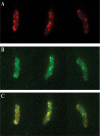
GFP-EpsM and mCherry-EpsC colocalization in V. cholerae cells. Cells of PBAD::eps producing both mCherry-EpsC and GFP-EpsM were imaged with DsRed (A) and GFP (B) filters. (C) Overlays of DsRed and GFP signals are shown, with GFP-EpsM in green, mCherry-EpsC in red, and overlapping signals in yellow.

References

    1. Bitter, W., M. Koster, M. Latijnhouwers, H. de Cock, and J. Tommassen. 1998. Formation of oligomeric rings by XcpQ and PilQ, which are involved in protein transport across the outer membrane of Pseudomonas aeruginosa. Mol. Microbiol. 27209-219. - PubMed
    1. Bleves, S., M. Gerard-Vincent, A. Lazdunski, and A. Filloux. 1999. Structure-function analysis of XcpP, a component involved in general secretory pathway-dependent protein secretion in Pseudomonas aeruginosa. J. Bacteriol. 1814012-4019. - PMC - PubMed
    1. Bouley, J., G. Condemine, and V. E. Shevchik. 2001. The PDZ domain of OutC and the N-terminal region of OutD determine the secretion specificity of the type II out pathway of Erwinia chrysanthemi. J. Mol. Biol. 308205-219. - PubMed
    1. Buddelmeijer, N., O. Francetic, and A. P. Pugsley. 2006. Green fluorescent chimeras indicate nonpolar localization of pullulanase secreton components PulL and PulM. J. Bacteriol. 1882928-2935. - PMC - PubMed
    1. Buddelmeijer, N., M. Krehenbrink, F. Pecorari, and A. P. Pugsley. 2009. Type II secretion system secretin PulD localizes in clusters in the Escherichia coli outer membrane. J. Bacteriol. 191161-168. - PMC - PubMed

Publication types

MeSH terms