. 2009 Jan 15;28(2):219-30.
doi: 10.1038/onc.2008.379. Epub 2008 Oct 6.

Rbpj conditional knockout reveals distinct functions of Notch4/Int3 in mammary gland development and tumorigenesis

Affiliations

Rbpj conditional knockout reveals distinct functions of Notch4/Int3 in mammary gland development and tumorigenesis

A Raafat et al. Oncogene. .

Abstract

Transgenic mice expressing the Notch 4 intracellular domain (ICD) (Int3) in the mammary gland have two phenotypes: arrest of mammary alveolar/lobular development and mammary tumorigenesis. Notch4 signaling is mediated primarily through the interaction of Int3 with the transcription repressor/activator Rbpj. We have conditionally ablated the Rbpj gene in the mammary glands of mice expressing whey acidic protein (Wap)-Int3. Interestingly, Rbpj knockout mice (Wap-Cre(+)/Rbpj(-/-)/Wap-Int3) have normal mammary gland development, suggesting that the effect of endogenous Notch signaling on mammary gland development is complete by day 15 of pregnancy. RBP-J heterozygous (Wap-Cre(+)/Rbpj(-/+)/Wap-Int3) and Rbpj control (Rbpj(flox/flox)/Wap-Int3) mice are phenotypically the same as Wap-Int3 mice with respect to mammary gland development and tumorigenesis. In addition, the Wap-Cre(+)/Rbpj(-/-)/Wap-Int3-knockout mice also developed mammary tumors at a frequency similar to Rbpj heterozygous and Wap-Int3 control mice but with a slightly longer latency. Thus, the effect on mammary gland development is dependent on the interaction of the Notch ICD with the transcription repressor/activator Rbpj, and Notch-induced mammary tumor development is independent of this interaction.


Figures

Figure 1
Figure 1
Genotyping and analysis of mice. Wap-Cre/Rbpjflox/flox mice were genetically crossed with Wap-Cre/ Rbpj+/flox/Wap-Int3 mice, to generate Wap-Cre+/Rbpjflox/flox/Wap-Int3 (Wap-Int3/Rbpj knockout), Wap-Cre+/Rbpjflox/+/Wap-Int3 (Wap-Int3/Rbpj heterozygous) and Rbpjflox/flox/Wap-Int3 (Wap-Int3/Rbpj control) mice, as described in the Materials and methods. (a) PCR tail DNA analysis of Wap-Cre/Rbpj −/−/Wap-Int3 (lane 1), Wap-Cre/Rbpj−/+/Wap-Int3 (lane 2) and Rbpjflox/flox/Wap-Int3 (lane 3, note that because of the large size of the neomycin phosphotransferase cassette inserted in Rbpj intron 7, Wap-Int3/Rbpj control mice test negative for Rbpj in the tail DNA analysis). (b) Neo RT–PCR analysis of total RNA extracted from three each of Wap-Int3/Rbpj knockout and Wap-Int3/Rbpj control tumors. Only the Wap-Int3/Rbpj knockout tumor RNAs are negative for Neo. RT–PCR, reverse transcriptase PCR.
Figure 2
Figure 2
Morphology, histology and immunohistochemical analysis of Wap-Int3/RBP-J knockout mammary glands. Photomicrographs of mammary gland wholemounts (ad); histological sections (eh); and β-casein immunohistochemistry (il) in the Wap-Int3/ Rbpj control (panels a, e, and i), Wap-Int3/Rbpj heterozygous (panels b, f and j), Wap-Int3/Rbpj knockout (panels c, g and k) and FVB (d, h and l) mice collected from the number 4 inguinal mammary gland from day 1 lactating mice. Of the experimental mice, only the Wap-Int3/Rbpj knockout mice were able to lactate, showed normal alveolar development (c, g) and were positive for the milk protein, β-casein (k), confirming the morphological and histological observations. Panels ah are at ×10 original magnification and panels il are at ×40 original magnification. Each treatment group contained at least 10 mice.
Figure 3
Figure 3
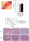
Mammary gland lesions, overall tumor-free survival and tumor histological analysis. (a) A representative wholemount from Wap-Int3/Rbpj knockout mouse showing several hyperproliferative lesions in the mammary gland; (b) the frequency of hyperproliferative lesions in Wap-Int3/Rbpj knockout, Wap-Int3/Rbpj heterozygous and Wap-Int3/Rbpj control mice in the fourth inguinal mammary gland, after the second parity; (c) the overall tumor-free survival of Wap-Int3/Rbpj control, Wap-Int3/Rbpj heterozygous and Wap-Int3/Rbpj knockout mice; histopathology of solid mammary adenocarcinomas from (d) Wap-Int3/Rbpj knockout tumor, (e) Wap-Int3/Rbpj heterozygous tumor, and (f) Wap-Int3/Rbpj control tumor; and (g) a papillary adenocarcinoma from Wap-Int3/Rbpj knockout mouse, (h) a Wap-Int3/Rbpj heterozygous tumor and (i) a Wap-Int3/Rbpj control tumor. All figures were hematoxylin-and-eosin-stained and are at ×40 original magnification.
Figure 4
Figure 4
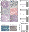
Photomicrographs of immunohistochemical staining of Neo and Rbpj in Wap-Int3/Rbpj knockout hyperplasia, primary mammary tumor and transplanted mammary tumor. (A) IHC analysis of Neo in Wap-Int3/Rbpj knockout (a, c and e) and Wap-Int3/ Rbpj control (b, d and f) hyperplasia (a, b), primary tumor (c, d) and tumor transplants (e, f). Only Wap-Int3/Rbpj control tissue was positive for Neo. Positive cells were scored in the hyperplasia (g), tumor (h) and mammary transplants (i) and labeling index was expressed as a percentage of positive nuclei of 3000 counted cells. In all tissues, neomycin phosphotransferse was significantly lower in Rbpj−/−/Wap-Int3 than Rbpj−/+/Wap-Int3. (B) IHC analysis of Rbpj using an antibody that recognizes the C′-terminal end of the protein (that is, wild-type protein) in a Wap-Int3/Rbpj knockout tumor (a) and Wap-Int3/Rbpj control tumor (b). Positive cells were scored in each and labeling index expressed as a percentage of positive nuclei of 3000 counted cells (c). A total of at least 5–6 mice were used for each experiment. *P<0.05. Original magnification was at ×40. IHC, immunohistochemistry.
Figure 5
Figure 5
Photomicrographs of immunohistochemical staining of Hes1 and Hey2 in Wap-Int3/Rbpj knockout hyperplasia, primary mammary tumor and transplanted mammary tumor. (A) IHC analysis of Hes-1 in Wap-Int3/Rbpj knockout (a, c and e) and Wap-Int3/ Rbpj control (b, d and f) hyperplasia (a, b), primary tumor (c, d) and transplanted tumor (e, f). Only Wap-Int3/Rbpj control tissue was positive for Hes-1. Positive cells were scored in the hyperplasia (g), tumor (h) and transplanted tumor (i) and labeling index was expressed as a percentage of positive nuclei of at least 3000 counted cells. In all tissues, Hes-1 was significantly lower in Wap-Int3/Rbpj knockout than Wap-Int3/Rbpj control. (B) IHC analysis of Hey2 in a Wap-Int3/Rbpj knockout tumor (a) and Wap-Int3/Rbpj control tumor (b). Positive cells were scored in each, and labeling index was expressed as a percentage of positive nuclei of 3000 counted cells (c). Hey2 was detected only in the Wap-Int3/Rbpj control tumor. A total of 5–6 mice were used for each experiment. *P<0.05. Original magnification was at ×40. IHC, immunohistochemistry.
Figure 6
Figure 6
In vivo effect of Rbpj deletion on proliferation and apoptosis of WAP-Int3/Rbpj knockout and control mammary tumors. WAP-Int3/Rbpj knockout (a and d) and control (b and e) tumor-bearing mice were euthanized and mammary tumor tissue was collected and processed for proliferation (a and b) and apoptosis (d and e) as described in the Materials and methods. Deletion of Rbpj in the WAP-Int3 mice did not affect the tumor proliferation; however, apoptosis was significantly higher in the WAP-Int3/Rbpj knockout mammary tumors than in the WAP-Int3/Rbpj control tumors. The proliferating and apoptotic cells were scored, and labeling index was expressed as a percentage of positive nuclei of at least 3000 counted cells. A total of at least 5–6 mice were used for each experiment. Arrows point to apoptotic cells. *P<0.05. Original magnification was at ×40.
Figure 7
Figure 7
Is Rbpj necessary for anchorage-independent growth of HC11 and HC11-Int3 cells in soft agar? HC11 and HC11-Int3 cells stably expressing Rbpj shRNA (HC11-H5shRNA, HC11-H6shRNA, HC11-Int3-H5shRNA, and HC11-Int3-H6shRNA) were tested for the expression of Rbpj RNA by RT–PCR (a), effect of Rbpj shRNA on Notch signaling (b) and proliferation (c) and for their ability to grow in soft agar (d and e). (a) HC11 (lane 1), HC11+scrambled vector (lane 2), HC11+RbpjH5shRNA (lane 3), HC11+RbpjH6shRNA (lane 4), HC11-int3 (lane 5), HC11-Int3+scrambled vector (lane 6), HC11-Int3+RbpjH5shRNA (lane 7) and HC11-Int3+RbpjH6shRNA (lane 8). RbpjH5 and H6shRNA both blocked Rbpj expression (Lanes 3, 4, 7 and 8), but RbpjH6shRNA was more efficient. (b) Quantitative RT–PCR analysis of Hey2 mRNA in the HC-11 (lane 1), HC11+ scrambled vector (lane 2), HC11+scrambled vector+ Rbpj H6shRNA (lane 3), Hc11+Int3 (lane 4), Hc11+Int3+RbpjH6shRNA (lane 5). Rbpj H6shRNA blocked Notch signaling (lane 4 vs lane 5). (c) Growth curves for HC11, HC11-Int3 and HC11-Int3-H6shRNA mammary epithelial cells. The rate of proliferation of HC11-Int3 and HC11-Int3 cells stably expressing Rbpj H6shRNA was not significantly different. (d) 15 000 cells were seeded in soft agar in the presence and absence of Rbpj H6shRNA. HC11 (lane 1), HC11- Int3 (lane 2), HC11+scrambled vector (lane 3), HC11+Rbpj H6shRNA (lane 4) and HC11-Int3+scrambled vector (lane 5). HC11 cells did not acquire the ability to grow in soft agar in the absence of Rbpj (lanes 4 and 5). (e) Soft agar growth of 15 000 HC11-Int3 cells in the presence and absence of Rbpj H6shRNA. HC11 (lane 1), HC11-Int3 (lane 2), HC11-Int3+scrambled vector (lane 3), HC11- Int3+Rbpj H6shRNA (lane 4). HC11-Int3 cells did not lose the ability to grow in soft agar in the absence of Rbpj (Lane 4). RT–PCR, reverse transcriptase PCR.

References

    1. Bashyam MD. Understanding cancer metastasis: an urgent need for using differential gene expression analysis. Cancer. 2002;94:1821–1829. - PubMed
    1. Bray S, Furriols M. Notch pathway: making sense of suppressor of hairless. Curr Biol. 2001;11:R217–R221. - PubMed
    1. Buono KD, Robinson GW, Martin C, Shi S, Stanley P, Tanigaki K, et al. The canonical Notch/RBP-J signaling pathway controls the balance of cell lineages in mammary epithelium during pregnancy. Dev Biol. 2006;293:565–580. - PubMed
    1. Callahan R, Egan SE. Notch signaling in mammary development and oncogenesis. J Mammary Gland Biol Neoplasia. 2004;9:145–163. - PubMed
    1. Dumont E, Fuchs KP, Bommer G, Christoph B, Kremmer E, Kempkes B. Neoplastic transformation by Notch is independent of transcriptional activation by RBP-J signalling. Oncogene. 2000;19:556–561. - PubMed

Publication types

MeSH terms