. 2007 Jul;18(7):2592-602.
doi: 10.1091/mbc.e06-12-1149. Epub 2007 May 2.

Accumulation of polyadenylated mRNA, Pab1p, eIF4E, and eIF4G with P-bodies in Saccharomyces cerevisiae

Affiliations

Accumulation of polyadenylated mRNA, Pab1p, eIF4E, and eIF4G with P-bodies in Saccharomyces cerevisiae

Muriel Brengues et al. Mol Biol Cell. 2007 Jul.

Abstract

Recent experiments have shown that mRNAs can move between polysomes and P-bodies, which are aggregates of nontranslating mRNAs associated with translational repressors and the mRNA decapping machinery. The transitions between polysomes and P-bodies and how the poly(A) tail and the associated poly(A) binding protein 1 (Pab1p) may affect this process are unknown. Herein, we provide evidence that poly(A)(+) mRNAs can enter P-bodies in yeast. First, we show that both poly(A)(-) and poly(A)(+) mRNA become translationally repressed during glucose deprivation, where mRNAs accumulate in P-bodies. In addition, both poly(A)(+) transcripts and/or Pab1p can be detected in P-bodies during glucose deprivation and in stationary phase. Cells lacking Pab1p have enlarged P-bodies, suggesting that Pab1p plays a direct or indirect role in shifting the equilibrium of mRNAs away from P-bodies and into translation, perhaps by aiding in the assembly of a type of mRNP within P-bodies that is poised to reenter translation. Consistent with this latter possibility, we observed the translation initiation factors (eIF)4E and eIF4G in P-bodies at a low level during glucose deprivation and at high levels in stationary phase. Moreover, Pab1p exited P-bodies much faster than Dcp2p when stationary phase cells were given fresh nutrients. Together, these results suggest that polyadenylated mRNAs can enter P-bodies, and an mRNP complex including poly(A)(+) mRNA, Pab1p, eIF4E, and eIF4G2 may represent a transition state during the process of mRNAs exchanging between P-bodies and translation.


Figures

Figure 1.
Figure 1.
poly(A)+ and poly(A) mRNAs can be translationally repressed. WT cells expressing the plasmid MFA2pG under the control of a tetracycline promoter (Tet-Off MFA2pG) were grown in SC medium plus Glu, shifted for 10 min to SC with no glucose containing doxycycline (10 min −Glu). Polysomes profiles of the collected sucrose gradients are shown. Fraction 1 corresponds to the top of the gradient. Northern blots of polyacrylamide gels for the indicated mRNA are shown.
Figure 2.
Figure 2.
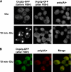
poly(A)+ mRNAs colocalize with P-bodies. WT cells expressing a GFP-tagged version of Dcp2p were grown in glucose containing medium (Glu) (A), shifted for 10 min without glucose (10 min −Glu) (A and B). Poly(A)+ RNA was visualized by oligo(dT) hybridization and indirect immunofluorescence. (B) Colocalization of poly(A)+ mRNAs with Dcp2-GFP. Left, Dcp2-GFP. Middle, poly(A)+. Right, merge generated by Adobe Photoshop (Adobe Systems, Mountain View, CA).
Figure 3.
Figure 3.
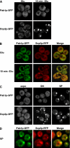
The poly(A) binding protein Pab1p is present in P-bodies under stress. WT cells expressing a GFP-tagged version of Dcp2p or Pab1p (A) or coexpressing a GFP-tagged version of Pab1p and a RFP-tagged version of Dcp2p (B) were grown in glucose-containing medium (Glu), shifted for 10 min without glucose (10 min −Glu). (B) Colocalization of Pab1p-GFP with Dcp2-RFP. Left, Pab1p-GFP. Middle, Dcp2p-RFP. Right, merge generated by Adobe Photoshop. (C) Cells expressing a GFP-tagged version of Dcp2p or Pab1p were observed at different stages of cellular growth, as described. (D) Colocalization of Pab1p-GFP with Dcp2-RFP during stationary phase.
Figure 4.
Figure 4.
Absence of Pab1p affects P-bodies distribution. WT, sbp2Δ, sbp2Δpab1Δ, pat1-2, and pat1-2pab1Δ cells expressing a GFP-tagged version of Dcp2p were grown in YPGlu.
Figure 5.
Figure 5.
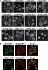
eIF4E and eIF4G2 are present in P-bodies during stationary phase. (A) Cells were grown in YPGlu (Glu) for 4 d. Cells expressing GFP-tagged versions of eIF1Ap, TIF35p, Prt1p, Nip1p, RPG1p, eIF4Bp, eIF4Ep, eIF4G1p, eIF4G2p, eIF5p eIF5Bp, and eIF6p are shown. (B) Colocalization of eIF4E-GFP and eIF4G2-GFP with Dcp2-RFP.
Figure 6.
Figure 6.
eIF4E, eIF4G1, and eIF4G2 are present in P-bodies under stress. (A) Cells were grown in YPGlu (Glu) and then shifted in YP without Glu for 10 min (10 min −Glu). Cells expressing GFP-tagged versions of eIF4Ep, eIF4G1p, eIF4G2p, and Dcp2p are shown. (B) Colocalization of eIF4E-GFP, eIF4G1-GFP, and eIF4G2-GFP with Dcp2-RFP under stress.
Figure 7.
Figure 7.
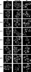
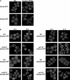
Effect of different factors on Pab1p accumulation in P-bodies. WT or mutant cells (as described) expressing a GFP-tagged version of Pab1p were grown in YPGlu (Glu) and then shifted in YP without Glu for 10 min (10 min −Glu) or were grown in SC medium plus Glu for 24 h and then shifted in YPGlu for three more days (SP).
Figure 8.
Figure 8.
Effect of translation initiation on Pab1p accumulation in P-bodies. (A) WT strain expressing a GFP-tagged version of Pab1p or Dcp2p grown in YPGlu (Glu) were exposed to 1 M KCl for 15 min. Cells were washed and resuspended in SC plus Glu supplemented or not with the same concentration of KCl and observed. (B) WT, cdc33–2 and prt1-63 strains expressing a GFP-tagged version of Pab1p or Dcp2p grown in SC medium plus glucose at 23°C were shifted to 39 or 37°C, respectively, for 1 h.
Figure 9.
Figure 9.
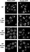
Pab1p exits P-bodies faster than Dcp2p. WT strains expressing a GFP-tagged version of Pab1p or Dcp2p were grown in YPGlu for 4 d (SP). Then, glucose was added to the culture, and the cells were observed at different time points as indicated.

References

    1. Ashe M. P., De Long S. K., Sachs A. B. Glucose depletion rapidly inhibits translation initiation in yeast. Mol. Biol. Cell. 2000;11:833–848. - PMC - PubMed
    1. Anderson J. S., Parker R. The 3′ to 5′ degradation of yeast mRNAs is a general mechanism for mRNA turnover that requires the SKI2 DEVH box protein and 3′ to 5′ exonucleases of the exosome complex. EMBO J. 1998;17:1497–1506. - PMC - PubMed
    1. Anderson P., Kedersha N. RNA granules. J. Cell Biol. 2006;172:803–808. - PMC - PubMed
    1. Barbee S. A., et al. Staufen and FMRP containing neuronal RNPs are structurally and functionally related to somatic P-bodies. Neuron. 2006;52:997–1009. - PMC - PubMed
    1. Beelman C. A., Parker R. Differential effects of translational inhibition in cis and in trans on the decay of the unstable yeast MFA2 mRNA. J. Biol. Chem. 1994;269:9687–9692. - PubMed

Publication types

MeSH terms