. 2007 Mar;189(5):1736-44.
doi: 10.1128/JB.01520-06. Epub 2006 Dec 8.

Dual negative control of spx transcription initiation from the P3 promoter by repressors PerR and YodB in Bacillus subtilis

Affiliations

Dual negative control of spx transcription initiation from the P3 promoter by repressors PerR and YodB in Bacillus subtilis

Montira Leelakriangsak et al. J Bacteriol. 2007 Mar.

Abstract

The spx gene encodes an RNA polymerase-binding protein that exerts negative and positive transcriptional control in response to oxidative stress in Bacillus subtilis. It resides in the yjbC-spx operon and is transcribed from at least five promoters located in the yjbC regulatory region or in the yjbC-spx intergenic region. Induction of spx transcription in response to treatment with the thiol-specific oxidant diamide is the result of transcription initiation at the P(3) promoter located upstream of the spx coding sequence. Previous studies conducted elsewhere and analyses of transcription factor mutants using transformation array technology have uncovered two transcriptional repressors, PerR and YodB, that target the cis-acting negative control elements of the P(3) promoter. Expression of an spx-bgaB fusion carrying the P(3) promoter is elevated in a yodB or perR mutant, and an additive increase in expression was observed in a yodB perR double mutant. Primer extension analysis of spx RNA shows the same additive increase in P(3) transcript levels in yodB perR mutant cells. Purified YodB and PerR repress spx transcription in vitro when wild-type spx P(3) promoter DNA was used as a template. Point mutations at positions within the P(3) promoter relieved YodB-dependent repression, while a point mutation at position +24 reduced PerR repression. DNase I footprinting analysis showed that YodB protects a region that includes the P(3) -10 and -35 regions, while PerR binds to a region downstream of the P(3) transcriptional start site. The binding of both repressors is impaired by the treatment of footprinting reactions with diamide or hydrogen peroxide. The study has uncovered a mechanism of dual negative control that relates to the oxidative stress response of gram-positive bacteria.


Figures

FIG. 1.
FIG. 1.
Transcription factor transformation array using an spx-lacZ fusion strain as a recipient. Colonies on the left are transformants of a strain that were transformed with DNA from individual mutants with an insertion in genes encoding known or putative transcription factors. The genes mutated are indicated on the right and correspond to the pattern of colonies on the left.
FIG. 2.
FIG. 2.
Effect of the disruption of perR, yodB, and perR yodB on the expression of spx-bgaB cells. The cells were grown in DSM, and their β-galactosidase (BgaB) activities were determined as described in Materials and Methods. Time zero indicates the mid-log phase. Triplicate experiments were performed. •, ORB6284 (wild type); ▪, ORB6288 (yodB mutant); ▴, ORB6268 (perR mutant); ×, ORB6594 (yodB perR double mutant). (B) Complementation experiment of yodB. β-Galactosidase activities of strains containing spx-bgaB and yodB::cat complemented with yodB are depicted. Cells were grown in DSM. Samples were taken after the OD600 reached 0.4 to 0.5 (time zero) and at 30 min, 60 min, 120 min, and 180 min. Results are means ± standard deviations from three independent experiments. •, ORB6284 (wild type); ▪, ORB6288 (yodB::cat); ▴, ORB6616 (yodB::cat thrC::yodB).
FIG. 3.
FIG. 3.
Primer extension analysis of RNA extracted from JH642, ORB6208 (yodB mutant), ORB6267 (perR mutant), and ORB6324 (yodB perR double mutant) cells in cultures subjected to diamide treatment. Cells were treated with 1 mM diamide for 10 min (10D) and without diamide (0 and 10 min) after the OD600 reached 0.4 to 0.5. Labeled primer oML02-15 was used for the primer extension reaction. The dideoxy sequencing ladders are shown on the left. For dideoxynucleotide sequencing, the nucleotide complementary to the dideoxynucleotide added in each reaction mixture is indicated above the corresponding lane (T′, A′, C′, and G′).
FIG. 4.
FIG. 4.
Runoff in vitro transcription analysis showed the effects of purified PerR and YodB on the levels of spx transcripts. Linear DNA templates for spx (deletions at positions +40 and +5 and point mutations T24C, T−26A, T−20G, T−19G, and A−14T) promoters and rpsD were generated by PCR. RNAP and DNA templates were incubated with or without PerR or YodB as indicated. The major transcript (bottom band) was quantified by using IMAGEQUANT data analysis software. The ratio of transcription (percent) was measured by comparing the transcripts from the reaction without repressor protein (as 100%) to transcripts with repressor protein for each template.
FIG. 5.
FIG. 5.
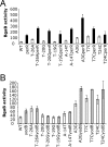
Effect of the perR (A) and yodB (B) disruption on the expression of spx promoter P3 point mutation cells. Cells were grown in DSM. The expression was determined as BgaB activity in Miller units at 30 min after cultures reached mid-log phase. Cells containing wild-type spx and point mutation (T−26A, T−20G, T−19G, A−14T, A3G, T7C, and T24C) promoters are indicated by white bars. Expression of the perR disruption and the perR disruption in spx point mutation cells is indicated by black bars (A). Expression of the yodB disruption and the yodB disruption in spx point mutation cells is indicated by gray bars (B). Results are means ± standard deviations from at least three independent experiments.
FIG. 6.
FIG. 6.
Result of DNase I footprinting of PerR and YodB to the top strand of the spx promoter. The wild-type spx promoter (A), point mutation T−26A spx promoter (B), point mutation T−19G spx promoter (C), and point mutation T+24C (lanes 1 to 3) and the wild type (lanes 4 to 6) (D) prepared by PCR were incubated in separate reaction mixtures with an increased amount of His-tagged PerR and YodB and subjected to DNase I cleavage. Lines in A indicate the protected regions, with a single line for PerR and double lines for YodB. The positions relative to the transcriptional start site are shown on the left (A and D). The nucleotide substitution (T24C) is indicated by an asterisk (D). BSA, bovine serum albumin.
FIG. 7.
FIG. 7.
Effect of diamide and hydrogen peroxide on DNA binding. (A) DNase I footprinting was used to assess the effect of diamide on YodB and PerR binding to the spx promoter (A) and on CymR binding to the yrrT promoter (B). The protected region by CymR is indicated by the dashed line on the right side. The positions relative to the transcriptional start site are shown. (C) Effect of H2O2 on DNA binding. PerR and YodB were incubated with DNA as follows: lanes 1 and 2, DNA-alone control; lanes 3 to 6, 11, and 12, 1 μM PerR; lanes 7 to 12, 2 μM YodB. The concentrations of H2O2 used were 25 mM, 50 mM, and 100 mM. BSA, bovine serum albumin.
FIG. 8.
FIG. 8.
Protection patterns from DNase I footprinting experiments. The nucleotide sequences of the spx promoter are shown. The regions protected by YodB (double solid lines) and by PerR (single solid line) are indicated. Putative −10 and −35 sequences are boxed. TSS, transcription start site. RBS, ribosome-binding site.

References

    1. Antelmann, H., C. Scharf, and M. Hecker. 2000. Phosphate starvation-inducible proteins of Bacillus subtilis: proteomics and transcriptional analysis. J. Bacteriol. 182:4478-4490. - PMC - PubMed
    1. Chatterjee, S. S., H. Hossain, S. Otten, C. Kuenne, K. Kuchmina, S. Machata, E. Domann, T. Chakraborty, and T. Hain. 2006. Intracellular gene expression profile of Listeria monocytogenes. Infect. Immun. 74:1323-1338. - PMC - PubMed
    1. Choi, S. Y., D. Reyes, M. Leelakriangsak, and P. Zuber. 2006. The global regulator Spx functions in the control of organosulfur metabolism in Bacillus subtilis. J. Bacteriol. 188:5741-5751. - PMC - PubMed
    1. Duwat, P., S. D. Ehrlich, and A. Gruss. 1999. Effects of metabolic flux on stress response pathways in Lactococcus lactis. Mol. Microbiol. 31:845-858. - PubMed
    1. Even, S., P. Burguiere, S. Auger, O. Soutourina, A. Danchin, and I. Martin-Verstraete. 2006. Global control of cysteine metabolism by CymR in Bacillus subtilis. J. Bacteriol. 188:2184-2197. - PMC - PubMed

Publication types

LinkOut - more resources