. 2006 Jun;18(6):1454-66.
doi: 10.1105/tpc.105.038695. Epub 2006 May 5.

One of two alb3 proteins is essential for the assembly of the photosystems and for cell survival in Chlamydomonas

Affiliations

One of two alb3 proteins is essential for the assembly of the photosystems and for cell survival in Chlamydomonas

Vera Göhre et al. Plant Cell. 2006 Jun.

Abstract

Proteins of the YidC/Oxa1p/ALB3 family play an important role in inserting proteins into membranes of mitochondria, bacteria, and chloroplasts. In Chlamydomonas reinhardtii, one member of this family, Albino3.1 (Alb3.1), was previously shown to be mainly involved in the assembly of the light-harvesting complex. Here, we show that a second member, Alb3.2, is located in the thylakoid membrane, where it is associated with large molecular weight complexes. Coimmunoprecipitation experiments indicate that Alb3.2 interacts with Alb3.1 and the reaction center polypeptides of photosystem I and II as well as with VIPP1, which is involved in thylakoid formation. Moreover, depletion of Alb3.2 by RNA interference to 25 to 40% of wild-type levels leads to a reduction in photosystems I and II, indicating that the level of Alb3.2 is limiting for the assembly and/or maintenance of these complexes in the thylakoid membrane. Although the levels of several photosynthetic proteins are reduced under these conditions, other proteins are overproduced, such as VIPP1 and the chloroplast chaperone pair Hsp70/Cdj2. These changes are accompanied by a large increase in vacuolar size and, after a prolonged period, by cell death. We conclude that Alb3.2 is required directly or indirectly, through its impact on thylakoid protein biogenesis, for cell survival.


Figures
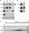
Figure 1.
Figure 1.
Localization of Alb3.2 Protein in the Chloroplast Compartment. (A) Immunoblot analysis of proteins from cw15 cells expressing FLAG-tagged Alb3.2. Extracts from purified chloroplasts (Chloro) and from chloroplast insoluble (C. ins) and soluble (C. sol) fractions were examined. The two bands in the immunoblot with Alb3.2 antiserum correspond to tagged (open arrowhead) and untagged (closed arrowhead) Alb3.2 protein. Samples of 20 μg of protein were loaded in each lane. (B) Immunoblot analysis of proteins from total cells and purified mitochondria (Mito) of cw15. (C) Alb3.2 is a thylakoid protein. Thylakoids from cw15 were separated from envelopes by centrifugation on a 0.1 to 2 M sucrose gradient for 4 h at 100,000g. Fractions were tested by immunoblotting. Filters were incubated with antisera raised against the indicated proteins: Alb3.2, PsaA, RbcS (small subunit of ribulose-1,5-bis-phosphate carboxylase/oxygenase), mCA (mitochondrial carbonic anhydrase), TrxH1 (thioredoxin H), D1, and ceQORH (chloroplast envelope quinone oxidoreductase homolog).
Figure 2.
Figure 2.
Alb3.1 and Alb3.2 Are Part of High Molecular Mass Complexes. (A) Thylakoid membranes from wild-type cells. The membranes were solubilized with β-dodecyl maltoside and fractionated by centrifugation on a linear 0.1 to 2 M sucrose gradient for 16 h at 170,000g. Fractions were used for immunoblotting with the indicated antibodies. (B) Thylakoid membranes from the ac29 strain containing HA-tagged Alb3.1 and FLAG-tagged Alb3.2. Membranes were solubilized and fractionated as described for (A). Fractions were used for immunoblotting with the HA, FLAG, D2, and PsaA antibodies. The lower Alb3.2 band is unspecific, as it is also observed with total protein extracts from an untagged wild-type strain (data not shown). Size markers are indicated.
Figure 3.
Figure 3.
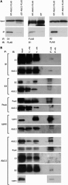
Alb3.2 Interacts with Alb3.1. Left, immunoprecipitations (IP) were performed with Alb3.2 antibodies (+Ab) or with preimmune serum (−Ab) with solubilized membranes of the ac29-Alb3.1-HA-Alb3.2-FLAG strain. Right, immunoprecipitations were performed with HA antibodies with solubilized membranes from the strains ac29-Alb3.1-HA-Alb3.2-FLAG (+HA) and ac29 (−HA). The immunoprecipitates were fractionated by PAGE, and immunoblotting (IB) was performed with the indicated antisera. Tagged and untagged Alb3.2 are indicated by open and closed arrowheads, respectively. f.t. refers to flow through.
Figure 4.
Figure 4.
Comigration of Alb3.2 with PSII Complexes on BN Gels. (A) Total membranes from the wild type and the mutant strains ac29, FuD7, F15, ΔpetD, and FuD50. The membranes were solubilized with β-dodecyl maltoside and fractionated by BN gel electrophoresis. The antibodies used for immunoblots are indicated at top. The arrow indicates the position of the presumed free Alb3.2. (B) Analysis of Alb3.2 in y-1. The y-1 mutant was grown in darkness and shifted to the light at 0 h. Total membranes were prepared at different times of the greening process and fractionated by BN gel electrophoresis (Gel). Immunoblotting was performed with Alb3.2 antiserum (Alb3.2). The location of the PSII core complex is indicated. CL, growth in continuous light. (C) Accumulation of proteins during greening of y-1. Total proteins from y-1 and the wild type were extracted during the greening process and fractionated by PAGE. Immunoblotting was performed with antisera raised against the indicated proteins. Rubisco, ribulose-1,5-bis-phosphate carboxylase/oxygenase.
Figure 5.
Figure 5.
Alb3.2 Interactions. (A) Alb3.2 interacts with D1 and D2. Immunoprecipitations like those described for Figure 3 were performed with extracts from the strain containing FLAG-tagged Alb3.2 and, as controls, the wild type and the strain ΔD1-Alb3.2-FLAG (FuD7 lacking psbA transformed with Alb3.2-FLAG). Input refers to an immunoblot (IB) of the extract before immunoprecipitation (IP) with the antiserum indicated for the immunoblot. (B) Alb3.2 interacts with PsaA and VIPP1. Similar immunoprecipitations of solubilized membranes of wild-type cells were performed with the indicated antisera (+Ab) or, as controls, with preimmune serum (−Ab). The input and flow through (f.t.) represent 10% of the proteins used for the immunoprecipitations. The immunoprecipitates were separated by PAGE and immunoblotted. Immunoblotting was performed with the indicated antisera.
Figure 6.
Figure 6.
Depletion of Alb3.2 by RNAi Leads to the Reduction of the Photosynthetic Complexes PSII and PSI. Wild-type cells were transformed with a plasmid carrying an inverted repeat of Alb3.2 cDNA using the cosilencing system described by Rohr et al. (2004). RNAi transformants were selected on 2.5 μM 5-FI and then transferred subsequently at 3-week intervals to 5, 10, 15, and 20 μM 5-FI. The transformants were always grown in the dark because 5-FI is light-sensitive. Total proteins (10 μg) from two independent transformants, Alb3.2_1 and Alb3.2_4, grown on 20 μM 5-FI for 2 weeks were used for PAGE and immunoblotted with several antibodies, as indicated. CDJ2, chloroplast DNAJ-like protein; VIPP1, vesicle-inducing protein. Serial dilutions from the FLP RNAi strain (control RNAi) (Falciatore et al., 2005) grown in the same conditions were used for quantification together with the wild type to correct for possible effects of 5-FI. Protein levels of Alb3.2_4 relative to the wild type are indicated at right. This experiment was repeated three times.
Figure 7.
Figure 7.
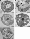
Depletion of Alb3.2 Leads to an Increase in Vacuolar Size. (A) Wild type (cw15). (B) RNAi strain Alb3.2_4. (C) RNAi strain Alb3.2_1. (D) RNAi control strain FLP. (E) Vacuole of Alb3.2_4 containing mitochondria. Cells were prepared for transmission electron microscopy as described in Methods. C, chloroplast; M, mitochondria; N, nucleus; V, vacuole. Bars = 1 μm.

References

    1. Barkan, A., and Goldschmidt-Clermont, M. (2000). Participation of nuclear genes in chloroplast gene expression. Biochimie 82 559–572. - PubMed
    1. Bartsevich, V.V., and Pakrasi, H.B. (1997). Molecular identification of a novel protein that regulates biogenesis of photosystem I, a membrane protein complex. J. Biol. Chem. 272 6382–6387. - PubMed
    1. Bellafiore, S., Ferris, P., Naver, H., Gohre, V., and Rochaix, J.D. (2002). Loss of Albino3 leads to the specific depletion of the light-harvesting system. Plant Cell 14 2303–2314. - PMC - PubMed
    1. Bonnefoy, N., Chalvet, F., Hamel, P., Slonimski, P.P., and Dujardin, G. (1994). OXA1, a Saccharomyces cerevisiae nuclear gene whose sequence is conserved from prokaryotes to eukaryotes controls cytochrome oxidase biogenesis. J. Mol. Biol. 239 201–212. - PubMed
    1. Boudreau, E., Takahashi, Y., Lemieux, C., Turmel, M., and Rochaix, J.D. (1997). The chloroplast ycf3 and ycf4 open reading frames of Chlamydomonas reinhardtii are required for the accumulation of the photosystem I complex. EMBO J. 16 6095–6104. - PMC - PubMed

Publication types

MeSH terms

Associated data

LinkOut - more resources