. 2001 Oct;21(19):6660-7.
doi: 10.1128/MCB.21.19.6660-6667.2001.

Recruitment of the class II phosphoinositide 3-kinase C2beta to the epidermal growth factor receptor: role of Grb2

Affiliations

Recruitment of the class II phosphoinositide 3-kinase C2beta to the epidermal growth factor receptor: role of Grb2

M Wheeler et al. Mol Cell Biol. 2001 Oct.

Abstract

Previously we demonstrated that the class II phosphoinositide 3-kinase C2beta (PI3K-C2beta) is rapidly recruited to a phosphotyrosine signaling complex containing the activated receptor for epidermal growth factor (EGF). Although this association was shown to be dependent upon specific phosphotyrosine residues present on the EGF receptor, the underlying mechanism remained unclear. In this study the interaction between PI3K-C2beta and the EGF receptor is competitively attenuated by synthetic peptides derived from each of three proline-rich motifs present within the N-terminal region of the PI3K. Further, a series of N-terminal PI3K-C2beta fragments, truncated prior to each proline-rich region, bound the receptor with decreased efficiency. A single proline-rich region was unable to mediate receptor association. Finally, an equivalent N-terminal fragment of PI3K-C2alpha that lacks similar proline-rich motifs was unable to affinity purify the activated EGF receptor from cell lysates. Since these findings revealed that the interaction between the EGF receptor and PI3K-C2beta is indirect, we sought to identify an adaptor molecule that could mediate their association. In addition to the EGF receptor, PI3K-C2beta(2-298) also isolated both Shc and Grb2 from A431 cell lysates. Recombinant Grb2 directly bound PI3K-C2beta in vitro, and this effect was reproduced using either SH3 domain expressed as a glutathione S-transferase (GST) fusion. Interaction with Grb2 dramatically increased the catalytic activity of this PI3K. The relevance of this association was confirmed when PI3K-C2beta was isolated by coimmunoprecipitation with anti-Grb2 antibody from numerous cell lines. Using immobilized, phosphorylated EGF receptor, recombinant PI3K-C2beta was only purified in the presence of Grb2. We conclude that proline-rich motifs within the N terminus of PI3K-C2beta mediate the association of this enzyme with activated EGF receptor and that this interaction involves the Grb2 adaptor.


Figures

FIG. 1
FIG. 1
The N terminus of PI3K-C2β but not PI3K-C2α interacts with the EGFR. A431 cells were incubated in the absence (−) or presence (+) of EGF (100 nM) for 10 min. Cell lysates were prepared and incubated with either GST, GST–PI3K-C2α(2-345), GST–PI3K-C2β(2-298), or GST–PI3K-C2α(1549-1686) for 4 h at 4°C. Fusion protein was isolated using glutathione-Sepharose beads, and associated proteins were fractionated by SDS-PAGE. Membranes were Western blotted with antiphosphotyrosine antibody (upper panel) or anti-EGFR antibody (lower panel). Arrows indicate the EGFR (approximately 180 kDa) and associated phosphoproteins. PY, phosphotyrosine.
FIG. 2
FIG. 2
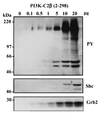
PI3K-C2β(2-298) affinity purifies EGFR, Shc, and Grb2 in a dose-dependent manner. Lysates from EGF-stimulated A431 cells were incubated with various additions of recombinant GST–PI3K-C2β(2-298) protein as shown. After 4 h at 4°C the fusion and associated proteins were isolated by centrifugation and were washed. They were then extracted, fractionated by SDS-PAGE, and Western blotted with antiphosphotyrosine (upper panel), anti-Shc (middle panel), or anti-Grb2 (lower panel). PY, phosphotyrosine.
FIG. 3
FIG. 3
Polyproline-rich peptides competitively attenuate the association of PI3K-C2β with the EGFR. Peptides (25 μM) corresponding to each of the three PI3K-C2β polyproline-rich regions at residues 127 to 140 (KKLSPPPLPPRASI), 140 to 153 (IWDTPPLPPRKGSP) and 252 to 265 (SKTMPPQVPPRTYA) or control residues 69 to 82 (NSLSPLEGPPNHST) were added to lysates of EGF-stimulated A431 cells. Samples were incubated at 4°C, and recombinant PI3K-C2β(2-298) (5 μg) was added 30 min later together with glutathione-Sepharose beads. After 4 h at 4°C, fusion protein was isolated by centrifugation, fractionated by SDS-PAGE, and Western blotted with antiphosphotyrosine (upper panel) or anti-PI3KC2β antibody (lower panel). PY, phosphotyrosine.
FIG. 4
FIG. 4
N-terminal fragments of PI3K-C2β establish that two proline-rich motifs are required for association with EGFR. A series of GST fusion proteins representing residues 2 to 130, 2 to 143, 2 to 157, 2 to 255, and 2 to 298 were incubated for 4 h at 4°C with lysates of A431 cells that had been treated in the absence (−) or presence (+) of EGF (100 nM) for 10 min. For control, EGFR was also immunoprecipitated from these lysates (Ab1; Oncogene Science). Each fusion and associated protein was isolated using glutathione-Sepharose beads, washed, fractionated by SDS-PAGE, and Western blotted with antiphosphotyrosine antibody to visualize the activated EGFR (upper panel) and anti-Grb2 antibody (lower panel). PY, phosphotyrosine.
FIG. 5
FIG. 5
Recombinant Grb2 and PI3K-C2β associate in vitro. Grb2-GST fusion protein (1 μg) was added to Triton lysis buffer (500 μl) in the absence or presence of EE epitope-tagged PI3K-C2β (1 μg) and either glutathione-Sepharose beads or anti-EE tag antibody (Ab) and protein A-Sepharose. After incubation at 4°C for 4 h, beads were isolated by centrifugation and washed. Associated proteins were extracted, fractionated by SDS-PAGE, and Western blotted with either anti-PI3K-C2β antibody (upper panel) or anti-Grb2 antibody (lower panel) and visualized with ECL.
FIG. 6
FIG. 6
Both N-terminal and C-terminal Grb2 SH3 domains (N-SH3 and C-SH3, respectively) bind PI3K-C2β. The N-terminal and C-terminal SH3 domains of Grb2 together with full-length Grb2 adaptor were expressed as GST fusion proteins and visualized by Coomassie blue staining following SDS-PAGE (panel A). The N-terminal PI3K-C2β fragments 2-130, 2-143, 2-157, 2-255, and 2-298 were expressed as GST fusion proteins, purified using glutathione-Sepharose beads, and cleaved with 5 μg of thrombin/ml (2 h, 21°C). Each protein was fractionated by SDS-PAGE and Western blotted with anti-PI3K-C2β antisera to confirm cross-reaction (panel B). Aliquots of each PI3K-C2β N-terminal fragment were incubated (2 h, 4°C) with either GST, full-length Grb2, or N-terminal or C-terminal Grb2 SH3 domain. Each GST fusion was isolated with glutathione-Sepharose beads, washed, and fractionated by SDS-PAGE. The N-terminal fragments of PI3K-C2β were visualized by Western blotting with anti-PI3K-C2β antibody.
FIG. 7
FIG. 7
Isolation of PI3K-C2β, SOS, and EGFR in anti-Grb2 immunoprecipitates (ippt). Confluent and quiescent cultures of human renal epithelial cells (HKC-8) and breast cancer cells (HTM3551, T47D, and SKBr3) were incubated with (+) or without (−) EGF (100 nM) for 10 min. Lysates were prepared to which anti-Grb2 antibody (Santa Cruz C-23) was added for 4 h at 4°C. Immune complexes were isolated following addition of protein A-Sepharose and centrifugation. Proteins were extracted, fractionated by SDS-PAGE, and Western blotted with antisera to PI3K-C2β (upper panels), SOS 1/2 (D21; Santa Cruz) (middle panels), and EGFR (1005; Santa Cruz) (lower panels).
FIG. 8
FIG. 8
Grb2 associates with PI3K-C2β in vivo. Cultures of confluent and quiescent A431, LNCaP, and HEK293 cells that overexpress epitope-tagged PI3K-C2β were incubated in the absence (−) or presence (+) of EGF (100 nM) for 10 min. Lysates were prepared and immunoprecipitated (ippt) with anti-Grb2 antisera for 4 h at 4°C. Immune complexes were isolated with protein A-Sepharose. These were either extracted, fractionated by SDS-PAGE, and Western blotted with anti-PI3K-C2β antisera (upper panels) or used for lipid kinase assay with PtdIns as the substrate and Ca2+ as the divalent cation (lower panels). The activity of recombinant PI3K-C2β is shown as a control, as is the class IA PI3K activity immunoprecipitated from A431 cell lysates using anti-p85α antibody.
FIG. 9
FIG. 9
Grb2 increases the catalytic activity of PI3K-C2β. Aliquots of recombinant EE-tagged PI3K-C2β (100 ng) were incubated in PI3K assay buffer for 2 h at 4°C with either Grb2-GST fusion protein (100 ng) or GST. After this time, immobilized EGFR previously immunoprecipitated (Ab-1; Oncogene Science) from lysates of quiescent A431 cells and autophosphorylated in vitro or mock control was added. Incubation was continued for a further 1 h. Following addition of kinase buffer, phosphatidylinositol (200 μg/ml) and [γ-32P]ATP, samples (50 μl) were incubated at room temperature for 20 min. Radiolabeled phosphoinositides were extracted and aliquots were fractionated by thin-layer chromatography and visualized by autoradiography. Representative data are shown in the upper panel. Quantification of PtdIns3P was undertaken by scanning densitometry. In the lower panel, data are displayed as means ± standard errors of the means (n = 6). Under the conditions used, all reactions displayed linear kinetics.
FIG. 10
FIG. 10
Formation of the EGFR-Grb2–PI3K-C2β complex in vitro. Recombinant EE-tagged PI3K-C2β (100 ng) was incubated in lysis buffer for 2 h at 4°C in the absence or presence of Grb2-GST fusion protein (100 ng) or GST. Immobilized EGFR, isolated by immunoprecipitation (Ab1; Oncogene Science) from lysates of confluent and quiescent cultures of A431 cells, was phosphorylated for 30 min at 30°C in protein kinase buffer upon addition of ATP. Either phosphorylated EGFR, nonphosphorylated EGFR, or mock immunoprecipitate was added to the Grb2–PI3K-C2β sample, and the incubation was continued for a further 1 h. Beads containing immobilized receptor and associated proteins were isolated by centrifugation, washed, fractionated by SDS-PAGE, and Western blotted with either anti-EGFR antibody, antiphosphotyrosine, anti-Grb2, or anti-PI3K-C2β antibody. PY, phosphotyrosine.

References

    1. Arcaro A, Volinia S, Zvelebil M J, Stein R, Watton S J, Layton M J, Gout I, Ahmadi K, Downward J, Waterfield M D. Human PI3-kinase C2β—the role of calcium and the C2 domain in enzyme activity. J Biol Chem. 1998;273:33082–33091. - PubMed
    1. Arcaro A, Zvelebil M J, Wallasch C, Ullrich A, Waterfield M D, Domin J. Class II phosphoinositide 3-kinase are downstream targets of activated polypeptide growth factor receptors. Mol Cell Biol. 2000;20:3817–3830. - PMC - PubMed
    1. Batzer A G, Blaikie P, Nelson K, Schlessinger J, Margolis B. The phosphotyrosine interaction domain of Shc binds an LXNPXY motif on the epidermal growth factor receptor. Mol Cell Biol. 1995;15:4403–4409. - PMC - PubMed
    1. Batzer A G, Rotin D, Urena J M, Skolnik E Y, Schlesinger J. Hierachy of binding sites for grb-2 and Shc on the epidermal growth factor receptor. Mol Cell Biol. 1994;14:5192–5201. - PMC - PubMed
    1. Brown R A, Domin J, Arcaro A, Waterfield M D, Shepherd P R. Insulin activates the alpha isoform of class II phosphoinositide 3-kinase. J Biol Chem. 1999;274:14529–14532. - PubMed

Publication types

MeSH terms