. 2001 Jan;21(2):644-54.
doi: 10.1128/MCB.21.2.644-654.2001.

Mitochondrial DNA instability and peri-implantation lethality associated with targeted disruption of nuclear respiratory factor 1 in mice

Affiliations

Mitochondrial DNA instability and peri-implantation lethality associated with targeted disruption of nuclear respiratory factor 1 in mice

L Huo et al. Mol Cell Biol. 2001 Jan.

Abstract

In vitro studies have implicated nuclear respiratory factor 1 (NRF-1) in the transcriptional expression of nuclear genes required for mitochondrial respiratory function, as well as for other fundamental cellular activities. We investigated here the in vivo function of NRF-1 in mammals by disrupting the gene in mice. A portion of the NRF-1 gene that encodes the nuclear localization signal and the DNA-binding and dimerization domains was replaced through homologous recombination by a beta-galactosidase-neomycin cassette. In the mutant allele, beta-galactosidase expression is under the control of the NRF-1 promoter. Embryos homozygous for NRF-1 disruption die between embryonic days 3.5 and 6.5. beta-Galactosidase staining was observed in growing oocytes and in 2. 5- and 3.5-day-old embryos, demonstrating that the NRF-1 gene is expressed during oogenesis and during early stages of embryogenesis. Moreover, the embryonic expression of NRF-1 did not result from maternal carryover. While most isolated wild-type and NRF-1(+/-) blastocysts can develop further in vitro, the NRF-1(-/-) blastocysts lack this ability despite their normal morphology. Interestingly, a fraction of the blastocysts from heterozygous matings had reduced staining intensity with rhodamine 123 and NRF-1(-/-) blastocysts had markedly reduced levels of mitochondrial DNA (mtDNA). The depletion of mtDNA did not coincide with nuclear DNA fragmentation, indicating that mtDNA loss was not associated with increased apoptosis. These results are consistent with a specific requirement for NRF-1 in the maintenance of mtDNA and respiratory chain function during early embryogenesis.


Figures

FIG. 1
FIG. 1
Targeted disruption of the mouse NRF-1 gene. (A) Schematic representation of the wild-type and the mutant mouse NRF-1 alleles covering CE1 to CE5 (filled boxes). The bold solid lines represent introns, and the thin solid lines indicate the plasmid backbone in the targeting vector. A promoterless β-galactosidase gene cassette (β-gal, hatched box) was inserted downstream of the first 33 bp of CE2 in the targeting vector. The neomycin cassette (neo, vertical hatched box) and the herpes simplex virus thymidine kinase cassette (hsv-tk, open box) each contain a mouse phosphoglycerate kinase promoter, and their transcriptional orientations are from right to left. The 5′ and 3′ homologous regions are 6 and 3.2 kb, respectively. Upon homologous recombination, the 5.3-kb β-gal-neo sequence replaces approximately 7 kb of endogenous NRF-1 gene sequence. Restriction enzyme cleavage sites shown above and below: B, BamHI; S, SacI; X, XbaI. The positions of probes used for genotyping by Southern blot analysis are indicated above the wild-type allele. Exons and the intron between CE4 and CE5 are not drawn to scale. (B) Southern blot analysis used to screen ES clones and genotype the progeny from heterozygous matings. The restriction enzymes and probes are shown above and below, respectively. The sizes of the hybridizing fragments in the wild-type (wt) and mutant (mut) alleles are shown on the right. (C) PCR primers for genotyping. The positions of the primers are indicated by solid bars. Primers 1 to 4 were designed to detect the wild-type allele, and primers 5 to 8 were designed to detect the mutant allele. In CE2, the sequence to the right of the dotted line is deleted in the mutant allele. Primers 1, 2, 5, and 6 are in the sense orientation, and primers 3, 4, 7, and 8 are in the antisense orientation. (D) PCR genotyping of the progeny from heterozygous matings. As described in Materials and Methods, newborn mice and 6.5- to 8.5-day-old embryos were genotyped with primers 2, 4, 5, and 7 shown in panel C, while preimplantation embryos were genotyped with primers 1 to 8 by a nested-PCR method. The sizes of the PCR products are indicated on the right of each panel.
FIG. 2
FIG. 2
β-Galactosidase expression. (A) Expression of NRF-1 and the NRF-1–β-galactosidase fusion gene transcripts in adult mice. A total of 10 μg of RNA isolated from the indicated tissues of an NRF-1+/− mouse (lanes 1 to 6) or from kidney of a wild-type littermate (lane 7) was analyzed for the expression of NRF-1 and NRF-1–β-galactosidase transcripts by RNase protection assay. Lane 6, with the NRF-1–β-galactosidase riboprobe alone; other lanes, with both NRF-1 and NRF-1–β-galactosidase riboprobe. The sizes of the protected products are indicated on the right. (B) β-Galactosidase expression in embryos. Embryonic stages are indicated on the left, and the various crosses are shown above. F, female; M, male. Positively stained embryos are indicated by arrows. β-Galactosidase activity was readily detected in eight-cell morulae (b and c) and in both the inner cell mass and trophectoderm cells in blastocysts (e and f).
FIG. 3
FIG. 3
Growth of blastocysts in culture. Blastocysts were cultured in vitro as described in Materials and Methods. (A) Blastocyst in culture medium immediately after isolation (day 0 in culture). (B and D) Blastocyst in culture for 40 h (day 2). (C and E) Blastocyst in culture for 100 h (day 5). Most blastocysts attached and started to grow at day 2 (B) and showed typical morphology at day 5, including the trophoblastic giant cells (TG) and the inner cell mass (ICM) (C). Putative NRF-1−/− blastocysts failed to grow (D and E). (B and C) The grown blastocyst was genotyped as NRF-1+/+. All of the images are at the same magnification.
FIG. 4
FIG. 4
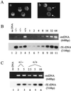
Mitochondrial staining and mtDNA copy number analyses. (A) Rhodamine 123 staining of blastocysts. Blastocysts were stained with rhodamine 123 as described in Materials and Methods. Four blastocysts from a wild-type mating (a) are compared to three blastocysts from a heterozygous mating (b). Those with weak fluorescence are indicated by the arrows. (B) mtDNA copy number in blastocysts. Half of the DNA isolated from each of the indicated embryos (lanes 2 to 4) was used to amplify a mtDNA fragment and a 5S rDNA fragment in a PCR. For standards, genomic DNA prepared from the heart of an adult wild-type mouse was diluted and subjected to the same PCR amplification (lanes 5 to 11). The amount of template was indicated in fold at the top for each standard. Products were resolved on a 1.2% agarose gel, and the mtDNA product was visualized by ethidium bromide staining. The 5S rDNA product was detected by Southern blotting. (C) mtDNA copy number in unfertilized eggs. Unfertilized eggs were isolated from wild-type or heterozygous NRF-1 females for DNA preparation as described in Materials and Methods. DNA from 5 eggs from a heterozygous animal (lanes 2 and 3) or from the equivalent of 2.5, 5, or 10 eggs from a wild-type animal (lanes 4, 5, and 6, respectively) was used as the template in PCR amplifications of the same mtDNA sequence as described in panel B. The numbers of eggs from which the template DNA was isolated are indicated above the panels.
FIG. 5
FIG. 5
NRF-1 expression in ovary. (A) β-Galactosidase staining of an ovary from a wild-type mouse. (B) β-Galactosidase staining of an ovary from a NRF-1+/− mouse. (C to F) β-Galactosidase staining of follicles from a NRF-1+/− mouse containing eggs at various stages of maturity. The corpus luteum (CL), the thecal cell layer surrounding the follicle (T), the follicle (Fl), the zona pellucida (ZP, unstained), the cytoplasm (Cy), and the nucleus (Nu) are indicated (B and E). The concentrated regions of β-galactosidase accumulation within the cytoplasm are indicated by arrows in panels C and F. Panels A and B are at the same magnification; panels C to F are at the same magnification.
FIG. 6
FIG. 6
Detection of chromosomal DNA fragmentation in blastocysts by TUNEL staining. TUNEL staining was performed on blastocysts as described in Materials and Methods. Blastocysts with expanded blastocoel cavities were selected to ensure that they had entered the stage when limited apoptosis would normally occur. Bright-field microscopy (A, C, and E) and the corresponding fluorescent images of TUNEL-stained blastocysts (B, D, and F) are shown. (A and B) Wild-type blastocyst treated with RQ1 DNase as a positive control. (C and D) Wild-type blastocyst showing very few stained cells. (E and F) NRF-1−/− blastocyst showing essentially the same staining intensity as the wild-type embryos.

References

    1. Abbondanzo S J, Gadi I, Stewart C L. Derivation of embryonic stem cell lines. Methods Enzymol. 1993;225:803–855. - PubMed
    1. Barbacci E, Reber M, Ott M-O, Breillat C, Huetz F, Cereghini S. Variant hepatocyte nuclear factor 1 is required for visceral endoderm specification. Development. 1999;126:4795–4805. - PubMed
    1. Becker T S, Burgess S M, Amsterdam A H, Allende M L, Hopkins N. not really finished is crucial for development of the zebrafish outer retina and encodes a transcription factor highly homologous to human nuclear respiratory factor 1 and avian initiation binding repressor. Development. 1998;124:4369–4378. - PubMed
    1. Bogarad L D, Arnone M I, Chang C, Davidson E H. Interference with gene regulation in living sea urchin embryos: transcription factor knock out (TKO), a genetically controlled vector for blockade of specific transcription factors. Proc Natl Acad Sci USA. 1998;95:14827–14832. - PMC - PubMed
    1. Carmeliet P, Lampugnani M G, Moons L, Breviario F, Compernolle V, Bono F, Balconi G, Spagnuolo R, Oostuyse B, Dewerchin M, Zanetti A, Angellilo A, Mattot V, Nuyens D, Lutgens E, Clotman F, de Ruiter M C, Gittenberger-de Groot A, Poelmann R, Lupu F, Herbert J M, Collen D, Dejana E. Targeted deficiency or cytosolic truncation of the VE-cadherin gene in mice impairs VEGF-mediated endothelial survival and angiogenesis. Cell. 1999;98:147–157. - PubMed

Publication types

MeSH terms

LinkOut - more resources